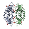
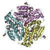
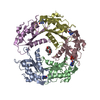
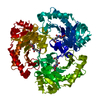
登録情報 データベース : PDB / ID : 1xn4タイトル PUTATIVE MAR1 RIBONUCLEASE FROM LEISHMANIA MAJOR ribonuclease MAR1 キーワード / / / / / 機能・相同性 / / / / / / / / 生物種 Leishmania major (大形リーシュマニア)手法 / / / 解像度 : 3.8 Å データ登録者 Caruthers, J. / Merritt, E.A. / Structural Genomics of Pathogenic Protozoa Consortium (SGPP) ジャーナル : Protein Sci. / 年 : 2005タイトル : Crystal structures and proposed structural/functional classification of three protozoan proteins from the isochorismatase superfamily.著者: Caruthers, J. / Zucker, F. / Worthey, E. / Myler, P.J. / Buckner, F. / Van Voorhuis, W. / Mehlin, C. / Boni, E. / Feist, T. / Luft, J. / Gulde, S. / Lauricella, A. / Kaluzhniy, O. / Anderson, ... 著者 : Caruthers, J. / Zucker, F. / Worthey, E. / Myler, P.J. / Buckner, F. / Van Voorhuis, W. / Mehlin, C. / Boni, E. / Feist, T. / Luft, J. / Gulde, S. / Lauricella, A. / Kaluzhniy, O. / Anderson, L. / Le Trong, I. / Holmes, M.A. / Earnest, T. / Soltis, M. / Hodgson, K.O. / Hol, W.G. / Merritt, E.A. 履歴 登録 2004年10月4日 登録サイト / 処理サイト 改定 1.0 2004年10月12日 Provider / タイプ 改定 1.1 2008年4月30日 Group 改定 1.2 2011年7月13日 Group / Derived calculations / Version format compliance改定 1.3 2012年10月24日 Group 改定 1.4 2023年8月23日 Group / Database references / Refinement descriptionカテゴリ chem_comp_atom / chem_comp_bond ... chem_comp_atom / chem_comp_bond / database_2 / pdbx_initial_refinement_model Item / _database_2.pdbx_database_accession
すべて表示 表示を減らす Remark 999 SEQUENCE This protein is a putative homologue of the MAR1 endoribonuclease from L tarentolae, SPTR ... SEQUENCE This protein is a putative homologue of the MAR1 endoribonuclease from L tarentolae, SPTR O77166. The closest Genbank sequence is the homologous protein from L Major, Genbank identifier LmjF12.0060.
 ムービー
ムービー コントローラー


 データを開く
 基本情報
基本情報 要素
要素 キーワード
キーワード 機能・相同性情報
 Leishmania major (大形リーシュマニア)
 X線回折 /
  データ登録者
 引用
引用 構造の表示
構造の表示 ダウンロードとリンク
 その他のダウンロード


 PDBj
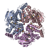
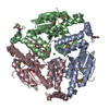
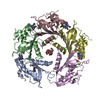
PDBj 集合体
集合体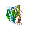


 試料調製
試料調製 / ビームライン: 8.2.2 / 波長: 0.9795
  解析
解析