 Movie
Movie Controller

[English] 日本語
 Yorodumi
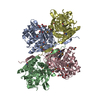
- PDB-1xl6: Intermediate gating structure 2 of the inwardly rectifying K+ cha... -
+ Open data

- Basic information
| Entry | Database: PDB / ID: 1xl6 | ||||||
|---|---|---|---|---|---|---|---|
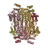
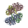
| Title | Intermediate gating structure 2 of the inwardly rectifying K+ channel KirBac3.1 | ||||||
 Components Components | Inward rectifier potassium channel | ||||||
 Keywords Keywords | METAL TRANSPORT / integral membrane protein / ion channel / inwardly rectifying K+ channel | ||||||
| Function / homology |  Function and homology information Function and homology informationregulation of monoatomic ion transmembrane transport / inward rectifier potassium channel activity / monoatomic ion channel complex / potassium ion import across plasma membrane / identical protein binding / plasma membrane Similarity search - Function | ||||||
| Biological species |  Magnetospirillum magnetotacticum (bacteria) Magnetospirillum magnetotacticum (bacteria) | ||||||
| Method |  X-RAY DIFFRACTION / MOLECULAR REPLACEMENT / Resolution: 2.85 Å X-RAY DIFFRACTION / MOLECULAR REPLACEMENT / Resolution: 2.85 Å | ||||||
 Authors Authors | Gulbis, J.M. / Kuo, A. / Smith, B. / Doyle, D.A. / Edwards, A. / Arrowsmith, C. / Sundstrom, M. | ||||||
 Citation Citation | Journal: To be Published Title: Two intermediate gating state crystal structures of the KirBac3.1 K+ channel Authors: Gulbis, J.M. / Kuo, A. / Smith, B. / Doyle, D.A. / Edwards, A. / Arrowsmith, C. / Sundstrom, M. | ||||||
| History |
|
- Structure visualization
| Structure viewer | Molecule: MolmilJmol/JSmol |
|---|
- Downloads & links
-Download
| PDBx/mmCIF format | 1xl6.cif.gz | 129.9 KB | Display | PDBx/mmCIF format |
|---|---|---|---|---|
| PDB format | pdb1xl6.ent.gz | 102.1 KB | Display | PDB format |
| PDBx/mmJSON format | 1xl6.json.gz | Tree view | PDBx/mmJSON format | |
| Others |  Other downloads Other downloads |
-Validation report
| Arichive directory | https://data.pdbj.org/pub/pdb/validation_reports/xl/1xl6ftp://data.pdbj.org/pub/pdb/validation_reports/xl/1xl6 | HTTPS FTP |
|---|
-Related structure data
| Related structure data |  1xl4C 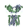 1p7bS C: citing same article ( S: Starting model for refinement |
|---|---|
| Similar structure data |
-Links
 PDBj
PDBj


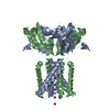
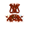
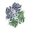
- Assembly
| Deposited unit | 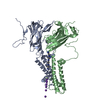
| |||||||||||||||||||||||||||||||||||||||||||||
|---|---|---|---|---|---|---|---|---|---|---|---|---|---|---|---|---|---|---|---|---|---|---|---|---|---|---|---|---|---|---|---|---|---|---|---|---|---|---|---|---|---|---|---|---|---|---|
| 1 | 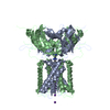
| |||||||||||||||||||||||||||||||||||||||||||||
| Unit cell |
| |||||||||||||||||||||||||||||||||||||||||||||
| Components on special symmetry positions |
|
-Components
| #1: Protein | Mass: 33782.766 Da / Num. of mol.: 2 Source method: isolated from a genetically manipulated source Source: (gene. exp.) Magnetospirillum magnetotacticum (bacteria)Plasmid: pET-30a / Production host: #2: Chemical | ChemComp-K /   Mass: 39.098 Da / Num. of mol.: 7 / Source method: obtained synthetically / Formula: K Mass: 39.098 Da / Num. of mol.: 7 / Source method: obtained synthetically / Formula: K#3: Chemical | ChemComp-MG / |   Mass: 24.305 Da / Num. of mol.: 1 / Source method: obtained synthetically / Formula: Mg Mass: 24.305 Da / Num. of mol.: 1 / Source method: obtained synthetically / Formula: Mg#4: Chemical | ChemComp-SPM / |   Mass: 202.340 Da / Num. of mol.: 1 / Source method: obtained synthetically / Formula: C10H26N4 Mass: 202.340 Da / Num. of mol.: 1 / Source method: obtained synthetically / Formula: C10H26N4#5: Water | ChemComp-HOH / |  Mass: 18.015 Da / Num. of mol.: 58 / Source method: isolated from a natural source / Formula: H2O Mass: 18.015 Da / Num. of mol.: 58 / Source method: isolated from a natural source / Formula: H2ONonpolymer details | THERE ARE THREE ALTERNATE ARRANGEMENTS OF IONS AND COORDINATED WATER MOLECULES. THEY ARE NOT ...THERE ARE THREE ALTERNATE ARRANGEMEN | |
|---|
-Experimental details
-Experiment
| Experiment | Method: X-RAY DIFFRACTION / Number of used crystals: 1 |
|---|
- Sample preparation
| Crystal | Density Matthews: 3.77 Å3/Da / Density % sol: 65 % |
|---|---|
| Crystal grow | Temperature: 293 K / Method: vapor diffusion, sitting drop / pH: 7.6 Details: 90mM Hepes, 2.5% PEG 8000, 2.5% PEG 4000, 20% PEG 400, 12.5mM magnesium chloride, 42.5mM magnesium acetate, 2.5% glycerol, 14mM Hega-10 , pH 7.6, VAPOR DIFFUSION, SITTING DROP, temperature 293K |
-Data collection
| Radiation | Protocol: SINGLE WAVELENGTH / Monochromatic (M) / Laue (L): M / Scattering type: x-ray |
|---|---|
| Radiation wavelength | Relative weight: 1 |
| Reflection | Resolution: 2.7→30 Å / Num. obs: 28510 |
- Processing
| Software |
| |||||||||||||||||||||
|---|---|---|---|---|---|---|---|---|---|---|---|---|---|---|---|---|---|---|---|---|---|---|
| Refinement | Method to determine structure: MOLECULAR REPLACEMENT Starting model: PDB ENTRY 1P7B Resolution: 2.85→10 Å / σ(F): 3
| |||||||||||||||||||||
| Refinement step | Cycle: LAST / Resolution: 2.85→10 Å
|
