 Movie
Movie Controller

[English] 日本語
 Yorodumi
- PDB-1xjx: The crystal structures of the DNA binding sites of the RUNX1 tran... -
+ Open data

- Basic information
| Entry | Database: PDB / ID: 1xjx | ||||||
|---|---|---|---|---|---|---|---|
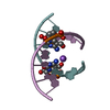
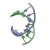
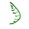
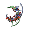
| Title | The crystal structures of the DNA binding sites of the RUNX1 transcription factor | ||||||
 Components Components |
| ||||||
 Keywords Keywords | DNA / A-DNA / DOUBLE HELIX / OVERHANGING BASES / RUNX1 | ||||||
| Function / homology | DNA Function and homology information Function and homology information | ||||||
| Method |  X-RAY DIFFRACTION / MOLECULAR REPLACEMENT / Resolution: 1.7 Å X-RAY DIFFRACTION / MOLECULAR REPLACEMENT / Resolution: 1.7 Å | ||||||
 Authors Authors | Kitayner, M. / Rozenberg, H. / Rabinovich, D. / Shakked, Z. | ||||||
 Citation Citation | Journal: Acta Crystallogr.,Sect.D / Year: 2005 Title: Structures of the DNA-binding site of Runt-domain transcription regulators. Authors: Kitayner, M. / Rozenberg, H. / Rabinovich, D. / Shakked, Z. | ||||||
| History |
|
- Structure visualization
| Structure viewer | Molecule: MolmilJmol/JSmol |
|---|
- Downloads & links
-Download
| PDBx/mmCIF format | 1xjx.cif.gz | 20.9 KB | Display | PDBx/mmCIF format |
|---|---|---|---|---|
| PDB format | pdb1xjx.ent.gz | 12.8 KB | Display | PDB format |
| PDBx/mmJSON format | 1xjx.json.gz | Tree view | PDBx/mmJSON format | |
| Others |  Other downloads Other downloads |
-Validation report
| Arichive directory | https://data.pdbj.org/pub/pdb/validation_reports/xj/1xjxftp://data.pdbj.org/pub/pdb/validation_reports/xj/1xjx | HTTPS FTP |
|---|
-Related structure data


-Links
 PDBj
PDBj

- Assembly
| Deposited unit | 
| ||||||||
|---|---|---|---|---|---|---|---|---|---|
| 1 |
| ||||||||
| Unit cell |
|
-Components
| #1: DNA chain | Mass: 2722.784 Da / Num. of mol.: 1 / Source method: obtained synthetically |
|---|---|
| #2: DNA chain | Mass: 2740.812 Da / Num. of mol.: 1 / Source method: obtained synthetically |
| #3: Water | ChemComp-HOH /  Mass: 18.015 Da / Num. of mol.: 102 / Source method: isolated from a natural source / Formula: H2O Mass: 18.015 Da / Num. of mol.: 102 / Source method: isolated from a natural source / Formula: H2O |
-Experimental details
-Experiment
| Experiment | Method: X-RAY DIFFRACTION / Number of used crystals: 1 |
|---|
- Sample preparation
| Crystal | Density Matthews: 1.83 Å3/Da / Density % sol: 32.92 % | ||||||||||||||||||||||||||||||||||||
|---|---|---|---|---|---|---|---|---|---|---|---|---|---|---|---|---|---|---|---|---|---|---|---|---|---|---|---|---|---|---|---|---|---|---|---|---|---|
| Crystal grow | Temperature: 293 K / Method: vapor diffusion, hanging drop / pH: 6.5 Details: MPD, magnesium chloride, sodium cacodylate acid, pH 6.5, VAPOR DIFFUSION, HANGING DROP, temperature 293K | ||||||||||||||||||||||||||||||||||||
| Components of the solutions |
|
-Data collection
| Diffraction | Mean temperature: 100 K | |||||||||
|---|---|---|---|---|---|---|---|---|---|---|
| Diffraction source | Source: ROTATING ANODE / Type: RIGAKU RUH3R / Wavelength: 1.54 / Wavelength: 1.5418 Å | |||||||||
| Detector | Type: RIGAKU RAXIS IV++ / Detector: IMAGE PLATE / Details: OSMIC CONFOCAL MIRROR | |||||||||
| Radiation | Protocol: SINGLE WAVELENGTH / Monochromatic (M) / Laue (L): M / Scattering type: x-ray | |||||||||
| Radiation wavelength |
| |||||||||
| Reflection | Resolution: 1.7→18.4 Å / Num. obs: 4315 / % possible obs: 95.9 % / Redundancy: 3.3 % / Rsym value: 0.039 / Net I/σ(I): 39.3 | |||||||||
| Reflection shell | Resolution: 1.7→1.73 Å / Redundancy: 1.9 % / Mean I/σ(I) obs: 4.9 / Rsym value: 0.236 / % possible all: 78.4 |
- Processing
| Software |
| |||||||||||||||||||||||||||||||||
|---|---|---|---|---|---|---|---|---|---|---|---|---|---|---|---|---|---|---|---|---|---|---|---|---|---|---|---|---|---|---|---|---|---|---|
| Refinement | Method to determine structure: MOLECULAR REPLACEMENT Starting model: FIBER A-DNA Resolution: 1.7→15 Å / Num. parameters: 1744 / Num. restraintsaints: 5767 / Cross valid method: THROUGHOUT / σ(F): 0 Stereochemistry target values: L.CLOWNEY ET AL.,J.AM.CHEM.SOC.118(1996)509-518, A.GELBIN ET AL.,J.AM.CHEM.SOC.118(1996)519-529, G.PARKINSON ET AL.,ACTA CRYST.D52(1996)57-64 Details: ANISOTROPIC SCALING APPLIED BY THE METHOD OF PARKIN, MOEZZI & HOPE, J.APPL.CRYST.28(1995)53-56
| |||||||||||||||||||||||||||||||||
| Refine analyze | Num. disordered residues: 0 / Occupancy sum hydrogen: 0 / Occupancy sum non hydrogen: 432 | |||||||||||||||||||||||||||||||||
| Refinement step | Cycle: LAST / Resolution: 1.7→15 Å
| |||||||||||||||||||||||||||||||||
| Refine LS restraints |
|
