 Movie
Movie Controller

[English] 日本語
 Yorodumi
- PDB-115d: ORDERED WATER STRUCTURE IN AN A-DNA OCTAMER AT 1.7 ANGSTROMS RESO... -
+ Open data

- Basic information
| Entry | Database: PDB / ID: 115d | ||||||||||||||||||
|---|---|---|---|---|---|---|---|---|---|---|---|---|---|---|---|---|---|---|---|
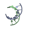
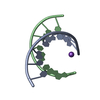
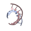
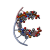
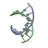
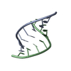
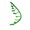
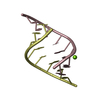
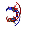
| Title | ORDERED WATER STRUCTURE IN AN A-DNA OCTAMER AT 1.7 ANGSTROMS RESOLUTION | ||||||||||||||||||
 Components Components | DNA (5'-D(* Keywords KeywordsDNA / A-DNA / DOUBLE HELIX / MODIFIED | Function / homology | DNA |  Function and homology information Function and homology informationMethod |  X-RAY DIFFRACTION / SYNCHROTRON / Resolution: 1.7 Å X-RAY DIFFRACTION / SYNCHROTRON / Resolution: 1.7 Å  Authors AuthorsKennard, O. / Cruse, W.B.T. / Nachman, J. / Prange, T. / Shakked, Z. / Rabinovich, D. |  CitationJournal: J.Biomol.Struct.Dyn. / Year: 1986 CitationJournal: J.Biomol.Struct.Dyn. / Year: 1986Title: Ordered water structure in an A-DNA octamer at 1.7 A resolution. Authors: Kennard, O. / Cruse, W.B. / Nachman, J. / Prange, T. / Shakked, Z. / Rabinovich, D. #1: Journal: Nature / Year: 1989Title: Coexistence of A-and B-Form DNA in a Single Crystal Lattice Authors: Doucet, J. / Benoit, J.-P. / Cruse, W.B.T. / Prange, T. / Kennard, O. #2: Journal: J.Mol.Biol. / Year: 1983Title: Sequence-Dependent Conformation of an A-DNA Double Helix. The Crystal Structure of the Octamer d(G-G-T-A-T-A-C-C) Authors: Shakked, Z. / Rabinovich, D. / Kennard, O. / Cruse, W.B.T. / Salisbury, S.A. / Viswamitra, M.A. #3: Journal: Proc.R.Soc.London,Ser.B / Year: 1981Title: Crystalline A-DNA. The X-Ray Analysis of the Fragment d(G-G-T-A-T-A-C-C) Authors: Shakked, Z. / Rabinovich, D. / Cruse, W.B.T. / Egert, E. / Kennard, O. / Sala, G. / Salisbury, S.A. / Viswamitra, M.A. History |
|
- Structure visualization
| Structure viewer | Molecule: MolmilJmol/JSmol |
|---|
- Downloads & links
-Download
| PDBx/mmCIF format | 115d.cif.gz | 18.6 KB | Display | PDBx/mmCIF format |
|---|---|---|---|---|
| PDB format | pdb115d.ent.gz | 12.2 KB | Display | PDB format |
| PDBx/mmJSON format | 115d.json.gz | Tree view | PDBx/mmJSON format | |
| Others |  Other downloads Other downloads |
-Validation report
| Arichive directory | https://data.pdbj.org/pub/pdb/validation_reports/15/115dftp://data.pdbj.org/pub/pdb/validation_reports/15/115d | HTTPS FTP |
|---|
-Related structure data
| Similar structure data |
|---|

-Links
 PDBj
PDBj

- Assembly
| Deposited unit | 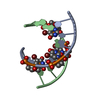
| ||||||||
|---|---|---|---|---|---|---|---|---|---|
| 1 |
| ||||||||
| Unit cell |
|
-Components
| #1: DNA chain | Mass: 2556.357 Da / Num. of mol.: 2 / Source method: obtained synthetically #2: Water | ChemComp-HOH / |  Mass: 18.015 Da / Num. of mol.: 85 / Source method: isolated from a natural source / Formula: H2O Mass: 18.015 Da / Num. of mol.: 85 / Source method: isolated from a natural source / Formula: H2O |
|---|
-Experimental details
-Experiment
| Experiment | Method: X-RAY DIFFRACTION |
|---|
- Sample preparation
| Crystal | Density Matthews: 2.39 Å3/Da / Density % sol: 48.62 % | |||||||||||||||||||||||||||||||||||
|---|---|---|---|---|---|---|---|---|---|---|---|---|---|---|---|---|---|---|---|---|---|---|---|---|---|---|---|---|---|---|---|---|---|---|---|---|
| Crystal grow | Method: vapor diffusion / pH: 7 / Details: pH 7.00, VAPOR DIFFUSION | |||||||||||||||||||||||||||||||||||
| Components of the solutions |
| |||||||||||||||||||||||||||||||||||
| Crystal grow | *PLUS pH: 7 | |||||||||||||||||||||||||||||||||||
| Components of the solutions | *PLUS
|
-Data collection
| Diffraction |
| ||||||||||||
|---|---|---|---|---|---|---|---|---|---|---|---|---|---|
| Diffraction source |
| ||||||||||||
| Detector |
| ||||||||||||
| Radiation |
| ||||||||||||
| Radiation wavelength |
| ||||||||||||
| Reflection | Highest resolution: 1.7 Å / Num. obs: 4669 / Observed criterion σ(F): 2 | ||||||||||||
| Reflection | *PLUS Highest resolution: 1.7 Å / Num. all: 5833 / Observed criterion σ(F): 2 / Rmerge(I) obs: 0.097 |

- Processing
| Software | Name: NUCLSQ / Classification: refinement | ||||||||||||
|---|---|---|---|---|---|---|---|---|---|---|---|---|---|
| Refinement | Resolution: 1.7→10 Å / σ(F): 2 /
| ||||||||||||
| Refine Biso |
| ||||||||||||
| Refinement step | Cycle: LAST / Resolution: 1.7→10 Å
| ||||||||||||
| Software | *PLUS Name: NUCLSQ / Classification: refinement | ||||||||||||
| Refinement | *PLUS Highest resolution: 1.7 Å / Lowest resolution: 10 Å / σ(F): 2 / Rfactor obs: 0.14 | ||||||||||||
| Solvent computation | *PLUS | ||||||||||||
| Displacement parameters | *PLUS | ||||||||||||
| Refine LS restraints | *PLUS
|
