 Movie
Movie Controller

[English] 日本語
 Yorodumi
- PDB-1xju: Crystal structure of secreted inactive form of P1 phage endolysin Lyz -
+ Open data

- Basic information
| Entry | Database: PDB / ID: 1xju | ||||||
|---|---|---|---|---|---|---|---|
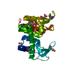
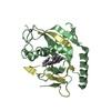
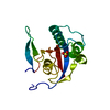
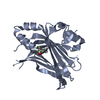
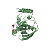
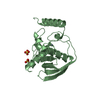
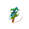
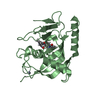
| Title | Crystal structure of secreted inactive form of P1 phage endolysin Lyz | ||||||
 Components Components | Lysozyme | ||||||
 Keywords Keywords | HYDROLASE / secreted inactive conformation | ||||||
| Function / homology |  Function and homology information Function and homology informationpeptidoglycan catabolic process / cell wall macromolecule catabolic process / lysozyme / lysozyme activity / killing of cells of another organism / defense response to bacterium / host cell plasma membrane Similarity search - Function | ||||||
| Biological species |  Enterobacteria phage P1 (virus) Enterobacteria phage P1 (virus) | ||||||
| Method |  X-RAY DIFFRACTION / SYNCHROTRON / MOLECULAR REPLACEMENT / Resolution: 1.07 Å X-RAY DIFFRACTION / SYNCHROTRON / MOLECULAR REPLACEMENT / Resolution: 1.07 Å | ||||||
 Authors Authors | Arockiasamy, A. / Sacchettini, J.C. | ||||||
 Citation Citation | Journal: Science / Year: 2005 Title: Disulfide isomerization after membrane release of its SAR domain activates P1 lysozyme. Authors: Xu, M. / Arulandu, A. / Struck, D.K. / Swanson, S. / Sacchettini, J.C. / Young, R. | ||||||
| History |
|
- Structure visualization
| Structure viewer | Molecule: MolmilJmol/JSmol |
|---|
- Downloads & links
-Download
| PDBx/mmCIF format | 1xju.cif.gz | 89.3 KB | Display | PDBx/mmCIF format |
|---|---|---|---|---|
| PDB format | pdb1xju.ent.gz | 66 KB | Display | PDB format |
| PDBx/mmJSON format | 1xju.json.gz | Tree view | PDBx/mmJSON format | |
| Others |  Other downloads Other downloads |
-Validation report
| Arichive directory | https://data.pdbj.org/pub/pdb/validation_reports/xj/1xjuftp://data.pdbj.org/pub/pdb/validation_reports/xj/1xju | HTTPS FTP |
|---|
-Related structure data


-Links
 PDBj
PDBj


- Assembly
| Deposited unit | 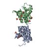
| ||||||||
|---|---|---|---|---|---|---|---|---|---|
| 1 | 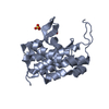
| ||||||||
| 2 | 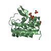
| ||||||||
| 3 | 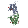
| ||||||||
| Unit cell |
|
-Components
| #1: Protein | Mass: 18478.969 Da / Num. of mol.: 2 Source method: isolated from a genetically manipulated source Source: (gene. exp.) Enterobacteria phage P1 (virus) / Genus: P1-like viruses / Gene: 17, LYSA, LyZ / Production host:  #2: Chemical | ChemComp-SO4 /   Mass: 96.063 Da / Num. of mol.: 8 / Source method: obtained synthetically / Formula: SO4 Mass: 96.063 Da / Num. of mol.: 8 / Source method: obtained synthetically / Formula: SO4#3: Water | ChemComp-HOH / |  Mass: 18.015 Da / Num. of mol.: 570 / Source method: isolated from a natural source / Formula: H2O Mass: 18.015 Da / Num. of mol.: 570 / Source method: isolated from a natural source / Formula: H2OHas protein modification | Y | |
|---|
-Experimental details
-Experiment
| Experiment | Method: X-RAY DIFFRACTION |
|---|
- Sample preparation
| Crystal | Density Matthews: 2.01 Å3/Da / Density % sol: 38.7 % |
|---|---|
| Crystal grow | Temperature: 291.15 K / Method: vapor diffusion / pH: 4.5 Details: sodium acetate, PEG4000, pH 4.5, VAPOR DIFFUSION, temperature 291.15K |
-Data collection
| Diffraction source | Source: SYNCHROTRON / Site: APS  / Beamline: 14-BM-C / Wavelength: 0.9 Å / Beamline: 14-BM-C / Wavelength: 0.9 Å | |||||||||||||||||||||||||||||||||||||||||||||||||||||||
|---|---|---|---|---|---|---|---|---|---|---|---|---|---|---|---|---|---|---|---|---|---|---|---|---|---|---|---|---|---|---|---|---|---|---|---|---|---|---|---|---|---|---|---|---|---|---|---|---|---|---|---|---|---|---|---|---|
| Radiation | Protocol: SINGLE WAVELENGTH / Monochromatic (M) / Laue (L): M / Scattering type: x-ray | |||||||||||||||||||||||||||||||||||||||||||||||||||||||
| Radiation wavelength | Wavelength: 0.9 Å / Relative weight: 1 | |||||||||||||||||||||||||||||||||||||||||||||||||||||||
| Reflection | Resolution: 1.07→50 Å / Num. obs: 116001 / % possible obs: 90.2 % / Redundancy: 5.6 % / Rmerge(I) obs: 0.057 | |||||||||||||||||||||||||||||||||||||||||||||||||||||||
| Reflection shell |
|
- Processing
| Software |
| ||||||||||||||||||||||||||||||||||||||||||||||||||||||||||||||||||||||||||||||||||||||||||||||||||||||||||||||||||||||||||||||||||||||||||||||||||||||||||||||||
|---|---|---|---|---|---|---|---|---|---|---|---|---|---|---|---|---|---|---|---|---|---|---|---|---|---|---|---|---|---|---|---|---|---|---|---|---|---|---|---|---|---|---|---|---|---|---|---|---|---|---|---|---|---|---|---|---|---|---|---|---|---|---|---|---|---|---|---|---|---|---|---|---|---|---|---|---|---|---|---|---|---|---|---|---|---|---|---|---|---|---|---|---|---|---|---|---|---|---|---|---|---|---|---|---|---|---|---|---|---|---|---|---|---|---|---|---|---|---|---|---|---|---|---|---|---|---|---|---|---|---|---|---|---|---|---|---|---|---|---|---|---|---|---|---|---|---|---|---|---|---|---|---|---|---|---|---|---|---|---|---|---|
| Refinement | Method to determine structure: MOLECULAR REPLACEMENT / Resolution: 1.07→46.62 Å / Cor.coef. Fo:Fc: 0.976 / Cor.coef. Fo:Fc free: 0.971 / Cross valid method: THROUGHOUT / σ(F): 0 / ESU R Free: 0.029 / Stereochemistry target values: Engh & Huber / Details: HYDROGENS HAVE BEEN ADDED IN THE RIDING POSITIONS
| ||||||||||||||||||||||||||||||||||||||||||||||||||||||||||||||||||||||||||||||||||||||||||||||||||||||||||||||||||||||||||||||||||||||||||||||||||||||||||||||||
| Solvent computation | Ion probe radii: 0.8 Å / Shrinkage radii: 0.8 Å / VDW probe radii: 1.2 Å / Solvent model: BABINET MODEL WITH MASK | ||||||||||||||||||||||||||||||||||||||||||||||||||||||||||||||||||||||||||||||||||||||||||||||||||||||||||||||||||||||||||||||||||||||||||||||||||||||||||||||||
| Displacement parameters | Biso mean: 9.67 Å2
| ||||||||||||||||||||||||||||||||||||||||||||||||||||||||||||||||||||||||||||||||||||||||||||||||||||||||||||||||||||||||||||||||||||||||||||||||||||||||||||||||
| Refinement step | Cycle: LAST / Resolution: 1.07→46.62 Å
| ||||||||||||||||||||||||||||||||||||||||||||||||||||||||||||||||||||||||||||||||||||||||||||||||||||||||||||||||||||||||||||||||||||||||||||||||||||||||||||||||
| Refine LS restraints |
| ||||||||||||||||||||||||||||||||||||||||||||||||||||||||||||||||||||||||||||||||||||||||||||||||||||||||||||||||||||||||||||||||||||||||||||||||||||||||||||||||
| LS refinement shell | Resolution: 1.07→1.098 Å / Total num. of bins used: 20 /
|
