 Movie
Movie Controller


+ Open data

- Basic information
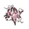
| Entry | Database: PDB / ID: 1xj6 | ||||||
|---|---|---|---|---|---|---|---|
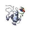
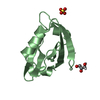
| Title | Structure of bjFixLH in the unliganded ferrous form | ||||||
 Components Components | Sensor protein fixL | ||||||
 Keywords Keywords | SIGNALING PROTEIN / TRANSFERASE / PAS domain / heme / oxygen sensor | ||||||
| Function / homology |  Function and homology information Function and homology informationhistidine phosphotransfer kinase activity / phosphorelay sensor kinase activity / histidine kinase / phosphorelay signal transduction system / regulation of DNA-templated transcription / ATP binding / metal ion binding / identical protein binding / plasma membrane Similarity search - Function | ||||||
| Biological species |  Bradyrhizobium japonicum (bacteria) Bradyrhizobium japonicum (bacteria) | ||||||
| Method |  X-RAY DIFFRACTION / SYNCHROTRON / MOLECULAR REPLACEMENT / Resolution: 1.9 Å X-RAY DIFFRACTION / SYNCHROTRON / MOLECULAR REPLACEMENT / Resolution: 1.9 Å | ||||||
 Authors Authors | Key, J. / Moffat, K. | ||||||
 Citation Citation | Journal: Biochemistry / Year: 2005 Title: Crystal Structures of Deoxy and CO-Bound bjFixLH Reveal Details of Ligand Recognition and Signaling Authors: Key, J. / Moffat, K. | ||||||
| History |
|
- Structure visualization
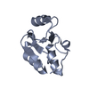
| Structure viewer | Molecule: MolmilJmol/JSmol |
|---|
- Downloads & links
-Download
| PDBx/mmCIF format | 1xj6.cif.gz | 61.1 KB | Display | PDBx/mmCIF format |
|---|---|---|---|---|
| PDB format | pdb1xj6.ent.gz | 43.9 KB | Display | PDB format |
| PDBx/mmJSON format | 1xj6.json.gz | Tree view | PDBx/mmJSON format | |
| Others |  Other downloads Other downloads |
-Validation report
| Arichive directory | https://data.pdbj.org/pub/pdb/validation_reports/xj/1xj6ftp://data.pdbj.org/pub/pdb/validation_reports/xj/1xj6 | HTTPS FTP |
|---|
-Related structure data
| Related structure data | 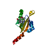 1xj2C 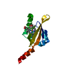 1xj3C 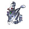 1xj4C 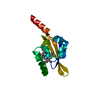 1drmS C: citing same article ( S: Starting model for refinement |
|---|---|
| Similar structure data |


-Links
 PDBj
PDBj


- Assembly
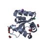
| Deposited unit | 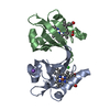
| ||||||||||||
|---|---|---|---|---|---|---|---|---|---|---|---|---|---|
| 1 | 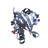
| ||||||||||||
| 2 | 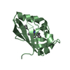
| ||||||||||||
| 3 | 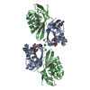
| ||||||||||||
| 4 | 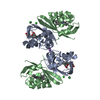
| ||||||||||||
| 5 |
| ||||||||||||
| 6 | 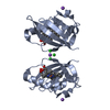
| ||||||||||||
| Unit cell |
| ||||||||||||
| Components on special symmetry positions |
|
-Components
| #1: Protein | Mass: 13413.079 Da / Num. of mol.: 2 / Fragment: heme domain Source method: isolated from a genetically manipulated source Details: unliganded / Source: (gene. exp.) Bradyrhizobium japonicum (bacteria) / Gene: fixL / Production host: References: UniProt: P23222, Transferases; Transferring phosphorus-containing groups; Phosphotransferases with a nitrogenous group as acceptor #2: Chemical |   Mass: 35.453 Da / Num. of mol.: 2 / Source method: obtained synthetically / Formula: Cl Mass: 35.453 Da / Num. of mol.: 2 / Source method: obtained synthetically / Formula: Cl#3: Chemical |   Mass: 22.990 Da / Num. of mol.: 2 / Source method: obtained synthetically / Formula: Na Mass: 22.990 Da / Num. of mol.: 2 / Source method: obtained synthetically / Formula: Na#4: Chemical |   Mass: 616.487 Da / Num. of mol.: 2 / Source method: obtained synthetically / Formula: C34H32FeN4O4 Mass: 616.487 Da / Num. of mol.: 2 / Source method: obtained synthetically / Formula: C34H32FeN4O4#5: Water | ChemComp-HOH / |  Mass: 18.015 Da / Num. of mol.: 102 / Source method: isolated from a natural source / Formula: H2O Mass: 18.015 Da / Num. of mol.: 102 / Source method: isolated from a natural source / Formula: H2O |
|---|
-Experimental details
-Experiment
| Experiment | Method: X-RAY DIFFRACTION / Number of used crystals: 2 |
|---|
- Sample preparation
| Crystal | Density Matthews: 2.51 Å3/Da / Density % sol: 43.4 % |
|---|---|
| Crystal grow | Temperature: 298 K / Method: vapor diffusion, hanging drop / pH: 8.6 Details: NaCl, Ethylene glycol, TRIS, pH 8.6, VAPOR DIFFUSION, HANGING DROP, temperature 298K |
-Data collection
| Diffraction | Mean temperature: 298 K |
|---|---|
| Diffraction source | Source: SYNCHROTRON / Site: APS  / Beamline: 14-BM-C / Wavelength: 0.9 Å / Beamline: 14-BM-C / Wavelength: 0.9 Å |
| Detector | Type: ADSC QUANTUM 4 / Detector: CCD |
| Radiation | Protocol: SINGLE WAVELENGTH / Monochromatic (M) / Laue (L): M / Scattering type: x-ray |
| Radiation wavelength | Wavelength: 0.9 Å / Relative weight: 1 |
| Reflection | Resolution: 1.9→40 Å / Num. all: 21079 / Num. obs: 19749 / % possible obs: 93.7 % / Observed criterion σ(I): -3 / Redundancy: 5.14 % / Rmerge(I) obs: 0.059 |
- Processing
| Software |
| |||||||||||||||||||||||||
|---|---|---|---|---|---|---|---|---|---|---|---|---|---|---|---|---|---|---|---|---|---|---|---|---|---|---|
| Refinement | Method to determine structure: MOLECULAR REPLACEMENT Starting model: 1DRM Resolution: 1.9→40 Å / Cross valid method: THROUGHOUT / σ(F): 2
| |||||||||||||||||||||||||
| Refinement step | Cycle: LAST / Resolution: 1.9→40 Å
| |||||||||||||||||||||||||
| Refine LS restraints |
|
