 Movie
Movie Controller

[English] 日本語
 Yorodumi
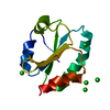
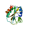
- PDB-3o6t: Mycobacterium tuberculosis thioredoxin C C40S mutant in Complex w... -
+ Open data

- Basic information
| Entry | Database: PDB / ID: 3o6t | ||||||
|---|---|---|---|---|---|---|---|
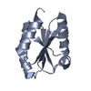
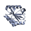
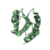
| Title | Mycobacterium tuberculosis thioredoxin C C40S mutant in Complex with Quinol Inhibitor PMX464 | ||||||
 Components Components | Thioredoxin | ||||||
 Keywords Keywords | ELECTRON TRANSPORT/INHIBITOR / Thioredoxin Fold / Electron Transport / ELECTRON TRANSPORT-INHIBITOR complex | ||||||
| Function / homology |  Function and homology information Function and homology informationCysteine synthesis from O-acetylserine / Cell redox homeostasis / Tolerance by Mtb to nitric oxide produced by macrophages / glutathione disulfide oxidoreductase activity / disulfide oxidoreductase activity / protein-disulfide reductase activity / cell redox homeostasis / peptidoglycan-based cell wall / electron transfer activity / extracellular region ...Cysteine synthesis from O-acetylserine / Cell redox homeostasis / Tolerance by Mtb to nitric oxide produced by macrophages / glutathione disulfide oxidoreductase activity / disulfide oxidoreductase activity / protein-disulfide reductase activity / cell redox homeostasis / peptidoglycan-based cell wall / electron transfer activity / extracellular region / plasma membrane / cytosol / cytoplasm Similarity search - Function | ||||||
| Biological species |   Mycobacterium tuberculosis (bacteria) Mycobacterium tuberculosis (bacteria) | ||||||
| Method |  X-RAY DIFFRACTION / SYNCHROTRON / MOLECULAR REPLACEMENT / Resolution: 2.4 Å X-RAY DIFFRACTION / SYNCHROTRON / MOLECULAR REPLACEMENT / Resolution: 2.4 Å | ||||||
 Authors Authors | Hall, G. / Emsley, J. | ||||||
 Citation Citation | Journal: Protein Sci. / Year: 2011 Title: Structure of Mycobacterium tuberculosis thioredoxin in complex with quinol inhibitor PMX464 Authors: Hall, G. / Bradshaw, T.D. / Laughton, C.A. / Stevens, M.F. / Emsley, J. | ||||||
| History |
|
- Structure visualization
| Structure viewer | Molecule: MolmilJmol/JSmol |
|---|
- Downloads & links
-Download
| PDBx/mmCIF format | 3o6t.cif.gz | 96.9 KB | Display | PDBx/mmCIF format |
|---|---|---|---|---|
| PDB format | pdb3o6t.ent.gz | 74.5 KB | Display | PDB format |
| PDBx/mmJSON format | 3o6t.json.gz | Tree view | PDBx/mmJSON format | |
| Others |  Other downloads Other downloads |
-Validation report
| Arichive directory | https://data.pdbj.org/pub/pdb/validation_reports/o6/3o6tftp://data.pdbj.org/pub/pdb/validation_reports/o6/3o6t | HTTPS FTP |
|---|
-Related structure data
| Related structure data |  3nofSC S: Starting model for refinement C: citing same article ( |
|---|---|
| Similar structure data |


-Links
 PDBj
PDBj


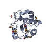
- Assembly
| Deposited unit | 
| ||||||||
|---|---|---|---|---|---|---|---|---|---|
| 1 | 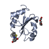
| ||||||||
| 2 | 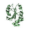
| ||||||||
| 3 | 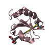
| ||||||||
| 4 | 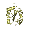
| ||||||||
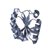
| Unit cell |
|
-Components
| #1: Protein | Mass: 12685.497 Da / Num. of mol.: 4 / Mutation: C40S Source method: isolated from a genetically manipulated source Source: (gene. exp.) Mycobacterium tuberculosis (bacteria) / Strain: H37Rv / Gene: TrxC / Plasmid: pGAT2 / Production host: #2: Chemical |   Mass: 243.281 Da / Num. of mol.: 2 / Source method: obtained synthetically / Formula: C13H9NO2S Mass: 243.281 Da / Num. of mol.: 2 / Source method: obtained synthetically / Formula: C13H9NO2S#3: Chemical |   Mass: 150.173 Da / Num. of mol.: 2 / Source method: obtained synthetically / Formula: C6H14O4 Mass: 150.173 Da / Num. of mol.: 2 / Source method: obtained synthetically / Formula: C6H14O4#4: Water | ChemComp-HOH / |  Mass: 18.015 Da / Num. of mol.: 149 / Source method: isolated from a natural source / Formula: H2O Mass: 18.015 Da / Num. of mol.: 149 / Source method: isolated from a natural source / Formula: H2OHas protein modification | Y | Nonpolymer details | THE LIGAND PX5 COVALENTLY BINDS TO CYS37 OF THIOREDOXIN THROUGH A MICHAEL ADDITION ONTO THE BETA- ...THE LIGAND PX5 COVALENTLY | |
|---|
-Experimental details
-Experiment
| Experiment | Method: X-RAY DIFFRACTION / Number of used crystals: 1 |
|---|
- Sample preparation
| Crystal | Density Matthews: 2.54 Å3/Da / Density % sol: 51.58 % |
|---|---|
| Crystal grow | Temperature: 292 K / Method: vapor diffusion, sitting drop / pH: 6.5 Details: 0.1M Na MES, pH 6.5, 4% PEG 400, 1.6M Ammonium Sulphate, VAPOR DIFFUSION, SITTING DROP, temperature 292K |
-Data collection
| Diffraction | Mean temperature: 93 K |
|---|---|
| Diffraction source | Source: SYNCHROTRON / Site: ESRF  / Beamline: ID29 / Wavelength: 0.976 Å / Beamline: ID29 / Wavelength: 0.976 Å |
| Detector | Type: ADSC QUANTUM 315r / Detector: CCD / Date: Sep 8, 2007 |
| Radiation | Monochromator: Si(111) / Protocol: SINGLE WAVELENGTH / Monochromatic (M) / Laue (L): M / Scattering type: x-ray |
| Radiation wavelength | Wavelength: 0.976 Å / Relative weight: 1 |
| Reflection | Resolution: 2.3→52.72 Å / Num. all: 22477 / Num. obs: 22477 / % possible obs: 97.7 % / Observed criterion σ(F): 1 / Observed criterion σ(I): 1 |
| Reflection shell | Resolution: 2.3→2.42 Å / Redundancy: 4.1 % / Rmerge(I) obs: 0.502 / Mean I/σ(I) obs: 2.8 / Num. unique all: 3304 / % possible all: 98.9 |
- Processing
| Software |
| ||||||||||||||||||||||||||||||||||||||||||||||||||||||||
|---|---|---|---|---|---|---|---|---|---|---|---|---|---|---|---|---|---|---|---|---|---|---|---|---|---|---|---|---|---|---|---|---|---|---|---|---|---|---|---|---|---|---|---|---|---|---|---|---|---|---|---|---|---|---|---|---|---|
| Refinement | Method to determine structure: MOLECULAR REPLACEMENT Starting model: PDB ENTRY 3NOF Resolution: 2.4→40.286 Å / SU ML: 0.35 / σ(F): 0 / Stereochemistry target values: ML
| ||||||||||||||||||||||||||||||||||||||||||||||||||||||||
| Solvent computation | Shrinkage radii: 0.9 Å / VDW probe radii: 1.11 Å / Solvent model: FLAT BULK SOLVENT MODEL / Bsol: 43.771 Å2 / ksol: 0.376 e/Å3 | ||||||||||||||||||||||||||||||||||||||||||||||||||||||||
| Displacement parameters |
| ||||||||||||||||||||||||||||||||||||||||||||||||||||||||
| Refinement step | Cycle: LAST / Resolution: 2.4→40.286 Å
| ||||||||||||||||||||||||||||||||||||||||||||||||||||||||
| Refine LS restraints |
| ||||||||||||||||||||||||||||||||||||||||||||||||||||||||
| LS refinement shell |
|
