 Movie
Movie Controller


+ Open data

- Basic information
| Entry | Database: PDB / ID: 1xfo | ||||||
|---|---|---|---|---|---|---|---|
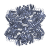
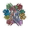
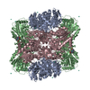
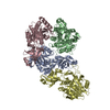
| Title | Crystal Structure of an archaeal aminopeptidase | ||||||
 Components Components | Frv operon protein FrvX | ||||||
 Keywords Keywords | HYDROLASE / aminopeptidase / self-compartmentalizing / metalloprotease / dinuclear | ||||||
| Function / homology |  Function and homology information Function and homology informationHydrolases; Acting on peptide bonds (peptidases); Aminopeptidases / aminopeptidase activity / metallopeptidase activity / proteolysis / metal ion binding Similarity search - Function | ||||||
| Biological species |   Pyrococcus horikoshii (archaea) Pyrococcus horikoshii (archaea) | ||||||
| Method |  X-RAY DIFFRACTION / SYNCHROTRON / MOLECULAR REPLACEMENT / Resolution: 1.96 Å X-RAY DIFFRACTION / SYNCHROTRON / MOLECULAR REPLACEMENT / Resolution: 1.96 Å | ||||||
 Authors Authors | Russo, S. / Baumann, U. | ||||||
 Citation Citation | Journal: J.Biol.Chem. / Year: 2004 Title: Crystal structure of a dodecameric tetrahedral shaped aminopeptidase Authors: Russo, S. / Baumann, U. | ||||||
| History |
|
- Structure visualization
| Structure viewer | Molecule: MolmilJmol/JSmol |
|---|
- Downloads & links
-Download
| PDBx/mmCIF format | 1xfo.cif.gz | 279.2 KB | Display | PDBx/mmCIF format |
|---|---|---|---|---|
| PDB format | pdb1xfo.ent.gz | 225.2 KB | Display | PDB format |
| PDBx/mmJSON format | 1xfo.json.gz | Tree view | PDBx/mmJSON format | |
| Others |  Other downloads Other downloads |
-Validation report
| Arichive directory | https://data.pdbj.org/pub/pdb/validation_reports/xf/1xfoftp://data.pdbj.org/pub/pdb/validation_reports/xf/1xfo | HTTPS FTP |
|---|
-Related structure data
| Related structure data | 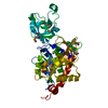 1vheS S: Starting model for refinement |
|---|---|
| Similar structure data |

-Links
 PDBj
PDBj- Assembly
| Deposited unit | 
| ||||||||||||||||||||||||||||||
|---|---|---|---|---|---|---|---|---|---|---|---|---|---|---|---|---|---|---|---|---|---|---|---|---|---|---|---|---|---|---|---|
| 1 | 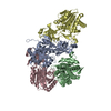
| ||||||||||||||||||||||||||||||
| Unit cell |
| ||||||||||||||||||||||||||||||
| Components on special symmetry positions |
| ||||||||||||||||||||||||||||||
| Noncrystallographic symmetry (NCS) | NCS domain:
NCS domain segments: Component-ID: 1 / Ens-ID: 1 / Beg auth comp-ID: MET / Beg label comp-ID: MET / End auth comp-ID: ILE / End label comp-ID: ILE / Refine code: 4 / Auth seq-ID: 6 - 353 / Label seq-ID: 10 - 357
| ||||||||||||||||||||||||||||||
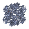
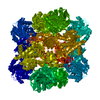
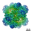
| Details | The physiological dodecamer is generated by the three fold axis at: 1/3, 2/3, z |
-Components
| #1: Protein | Mass: 39510.496 Da / Num. of mol.: 4 Source method: isolated from a genetically manipulated source Source: (gene. exp.) Pyrococcus horikoshii (archaea) / Plasmid: pET-28a / Production host:  #2: Chemical | ChemComp-ZN /   Mass: 65.409 Da / Num. of mol.: 8 / Source method: obtained synthetically / Formula: Zn Mass: 65.409 Da / Num. of mol.: 8 / Source method: obtained synthetically / Formula: Zn#3: Water | ChemComp-HOH / |  Mass: 18.015 Da / Num. of mol.: 645 / Source method: isolated from a natural source / Formula: H2O Mass: 18.015 Da / Num. of mol.: 645 / Source method: isolated from a natural source / Formula: H2O |
|---|
-Experimental details
-Experiment
| Experiment | Method: X-RAY DIFFRACTION / Number of used crystals: 1 |
|---|
- Sample preparation
| Crystal | Density Matthews: 2.6 Å3/Da / Density % sol: 51.7 % |
|---|---|
| Crystal grow | Temperature: 293 K / Method: vapor diffusion, sitting drop / pH: 6 Details: PEG 400, sodium chloride, pH 6.0, VAPOR DIFFUSION, SITTING DROP, temperature 293K |
-Data collection
| Diffraction | Mean temperature: 100 K |
|---|---|
| Diffraction source | Source: SYNCHROTRON / Site: ESRF  / Beamline: ID14-2 / Wavelength: 0.933 Å / Beamline: ID14-2 / Wavelength: 0.933 Å |
| Detector | Type: MARRESEARCH / Detector: CCD / Date: Apr 11, 2004 / Details: mirrors |
| Radiation | Monochromator: mirrors / Protocol: SINGLE WAVELENGTH / Monochromatic (M) / Laue (L): M / Scattering type: x-ray |
| Radiation wavelength | Wavelength: 0.933 Å / Relative weight: 1 |
| Reflection | Resolution: 1.95→40 Å / Num. all: 110425 / Num. obs: 110425 / % possible obs: 100 % / Observed criterion σ(F): 0 / Observed criterion σ(I): -3 / Redundancy: 4.1 % / Rmerge(I) obs: 0.037 / Net I/σ(I): 24.3 |
| Reflection shell | Resolution: 1.95→2.07 Å / Rmerge(I) obs: 0.193 / Mean I/σ(I) obs: 5.2 / % possible all: 63.5 |
- Processing
| Software |
| |||||||||||||||||||||||||||||||||||||||||||||||||||||||||||||||||||||||||||||||||||||||||||||||||||||||||||||||||||||||||||||
|---|---|---|---|---|---|---|---|---|---|---|---|---|---|---|---|---|---|---|---|---|---|---|---|---|---|---|---|---|---|---|---|---|---|---|---|---|---|---|---|---|---|---|---|---|---|---|---|---|---|---|---|---|---|---|---|---|---|---|---|---|---|---|---|---|---|---|---|---|---|---|---|---|---|---|---|---|---|---|---|---|---|---|---|---|---|---|---|---|---|---|---|---|---|---|---|---|---|---|---|---|---|---|---|---|---|---|---|---|---|---|---|---|---|---|---|---|---|---|---|---|---|---|---|---|---|---|
| Refinement | Method to determine structure: MOLECULAR REPLACEMENT Starting model: 1VHE Resolution: 1.96→29.92 Å / Cor.coef. Fo:Fc: 0.958 / Cor.coef. Fo:Fc free: 0.948 / SU B: 6.51 / SU ML: 0.096 / TLS residual ADP flag: LIKELY RESIDUAL / Cross valid method: THROUGHOUT / σ(F): 0 / σ(I): 0 / ESU R: 0.151 / ESU R Free: 0.133 / Stereochemistry target values: MAXIMUM LIKELIHOOD
| |||||||||||||||||||||||||||||||||||||||||||||||||||||||||||||||||||||||||||||||||||||||||||||||||||||||||||||||||||||||||||||
| Solvent computation | Ion probe radii: 0.8 Å / Shrinkage radii: 0.8 Å / VDW probe radii: 1.4 Å / Solvent model: BABINET MODEL WITH MASK | |||||||||||||||||||||||||||||||||||||||||||||||||||||||||||||||||||||||||||||||||||||||||||||||||||||||||||||||||||||||||||||
| Displacement parameters | Biso mean: 37.662 Å2
| |||||||||||||||||||||||||||||||||||||||||||||||||||||||||||||||||||||||||||||||||||||||||||||||||||||||||||||||||||||||||||||
| Refinement step | Cycle: LAST / Resolution: 1.96→29.92 Å
| |||||||||||||||||||||||||||||||||||||||||||||||||||||||||||||||||||||||||||||||||||||||||||||||||||||||||||||||||||||||||||||
| Refine LS restraints |
| |||||||||||||||||||||||||||||||||||||||||||||||||||||||||||||||||||||||||||||||||||||||||||||||||||||||||||||||||||||||||||||
| Refine LS restraints NCS | Ens-ID: 1 / Number: 2610 / Refine-ID: X-RAY DIFFRACTION
| |||||||||||||||||||||||||||||||||||||||||||||||||||||||||||||||||||||||||||||||||||||||||||||||||||||||||||||||||||||||||||||
| LS refinement shell | Resolution: 1.958→1.998 Å / Total num. of bins used: 25
| |||||||||||||||||||||||||||||||||||||||||||||||||||||||||||||||||||||||||||||||||||||||||||||||||||||||||||||||||||||||||||||
| Refinement TLS params. | Method: refined / Refine-ID: X-RAY DIFFRACTION
| |||||||||||||||||||||||||||||||||||||||||||||||||||||||||||||||||||||||||||||||||||||||||||||||||||||||||||||||||||||||||||||
| Refinement TLS group |
|
