 Movie
Movie Controller

[English] 日本語
 Yorodumi
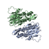
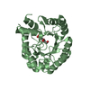
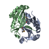
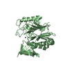
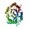
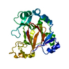
- PDB-1xfj: Crystal structure of protein CC_0490 from Caulobacter crescentus,... -
+ Open data

- Basic information
| Entry | Database: PDB / ID: 1xfj | ||||||
|---|---|---|---|---|---|---|---|
| Title | Crystal structure of protein CC_0490 from Caulobacter crescentus, Pfam DUF152 | ||||||
 Components Components | conserved hypothetical protein | ||||||
 Keywords Keywords | structural genomics / unknown function / Protein structure initiative (PSI) / Hypothetical protein / alpha-beta-beta-alpha / two-domain structure / New York SGX Research Center for Structural Genomics / NYSGXRC | ||||||
| Function / homology |  Function and homology information Function and homology informationS-methyl-5-thioadenosine phosphorylase activity / copper ion binding / hydrolase activity Similarity search - Function | ||||||
| Biological species |  Caulobacter vibrioides (bacteria) Caulobacter vibrioides (bacteria) | ||||||
| Method |  X-RAY DIFFRACTION / SYNCHROTRON / MAD / Resolution: 1.75 Å X-RAY DIFFRACTION / SYNCHROTRON / MAD / Resolution: 1.75 Å | ||||||
 Authors Authors | Krishnamurthy, N.R. / Kumaran, D. / Swaminathan, S. / Burley, S.K. / New York SGX Research Center for Structural Genomics (NYSGXRC) | ||||||
 Citation Citation | Journal: To be Published Title: Crystal structure of a conserved hypothetical protein from Caulobacter crescentus Authors: Krishnamurthy, N.R. / Kumaran, D. / Swaminathan, S. | ||||||
| History |
|
- Structure visualization
| Structure viewer | Molecule: MolmilJmol/JSmol |
|---|
- Downloads & links
-Download
| PDBx/mmCIF format | 1xfj.cif.gz | 68.4 KB | Display | PDBx/mmCIF format |
|---|---|---|---|---|
| PDB format | pdb1xfj.ent.gz | 48.8 KB | Display | PDB format |
| PDBx/mmJSON format | 1xfj.json.gz | Tree view | PDBx/mmJSON format | |
| Others |  Other downloads Other downloads |
-Validation report
| Arichive directory | https://data.pdbj.org/pub/pdb/validation_reports/xf/1xfjftp://data.pdbj.org/pub/pdb/validation_reports/xf/1xfj | HTTPS FTP |
|---|
-Related structure data
| Related structure data | |
|---|---|
| Similar structure data | |
| Other databases |


-Links
 PDBj
PDBj


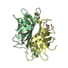
- Assembly
| Deposited unit | 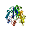
| ||||||||
|---|---|---|---|---|---|---|---|---|---|
| 1 |
| ||||||||
| Unit cell |
|
-Components
| #1: Protein | Mass: 28148.154 Da / Num. of mol.: 1 Source method: isolated from a genetically manipulated source Source: (gene. exp.) Caulobacter vibrioides (bacteria) / Production host: |
|---|---|
| #2: Chemical | ChemComp-ACT /   Mass: 59.044 Da / Num. of mol.: 1 / Source method: obtained synthetically / Formula: C2H3O2 Mass: 59.044 Da / Num. of mol.: 1 / Source method: obtained synthetically / Formula: C2H3O2 |
| #3: Chemical | ChemComp-BME /   Mass: 78.133 Da / Num. of mol.: 1 / Source method: obtained synthetically / Formula: C2H6OS Mass: 78.133 Da / Num. of mol.: 1 / Source method: obtained synthetically / Formula: C2H6OS |
| #4: Chemical | ChemComp-GOL /   Mass: 92.094 Da / Num. of mol.: 1 / Source method: obtained synthetically / Formula: C3H8O3 Mass: 92.094 Da / Num. of mol.: 1 / Source method: obtained synthetically / Formula: C3H8O3 |
| #5: Water | ChemComp-HOH /  Mass: 18.015 Da / Num. of mol.: 253 / Source method: isolated from a natural source / Formula: H2O Mass: 18.015 Da / Num. of mol.: 253 / Source method: isolated from a natural source / Formula: H2O |
| Has protein modification | Y |
-Experimental details
-Experiment
| Experiment | Method: X-RAY DIFFRACTION / Number of used crystals: 1 |
|---|
- Sample preparation
| Crystal | Density Matthews: 2 Å3/Da / Density % sol: 40 % |
|---|---|
| Crystal grow | Temperature: 293 K / Method: vapor diffusion, sitting drop / pH: 4.6 Details: PEG 4000, Sodium-acetate, pH 4.6, VAPOR DIFFUSION, SITTING DROP, temperature 293K |
-Data collection
| Diffraction | Mean temperature: 100 K | ||||||||||||
|---|---|---|---|---|---|---|---|---|---|---|---|---|---|
| Diffraction source | Source: SYNCHROTRON / Site: NSLS  / Beamline: X12C / Wavelength: 0.9792, 0.9797, 0.94 / Beamline: X12C / Wavelength: 0.9792, 0.9797, 0.94 | ||||||||||||
| Detector | Type: BRANDEIS - B4 / Detector: CCD / Date: Aug 14, 2004 / Details: mirrors | ||||||||||||
| Radiation | Monochromator: Si 111 CHANNEL / Protocol: MAD / Monochromatic (M) / Laue (L): M / Scattering type: x-ray | ||||||||||||
| Radiation wavelength |
| ||||||||||||
| Reflection | Resolution: 1.75→50 Å / Num. all: 21008 / Num. obs: 21008 / % possible obs: 97.8 % / Observed criterion σ(F): 0 / Observed criterion σ(I): 0 / Redundancy: 7.1 % / Biso Wilson estimate: 10 Å2 / Rmerge(I) obs: 0.052 / Net I/σ(I): 15.8 | ||||||||||||
| Reflection shell | Resolution: 1.75→1.81 Å / Redundancy: 4.1 % / Rmerge(I) obs: 0.125 / Num. unique all: 1760 / % possible all: 81.4 |
- Processing
| Software |
| ||||||||||||||||||||
|---|---|---|---|---|---|---|---|---|---|---|---|---|---|---|---|---|---|---|---|---|---|
| Refinement | Method to determine structure: MAD / Resolution: 1.75→50 Å / Cross valid method: THROUGHOUT / σ(F): 0 / Stereochemistry target values: Engh & Huber Details: Ser 38 and Asp 113 are in the disallowed region of the Ramachandran plot. This seems to be the characteristic feature of this protein. It is observed in the other related entries as well. ...Details: Ser 38 and Asp 113 are in the disallowed region of the Ramachandran plot. This seems to be the characteristic feature of this protein. It is observed in the other related entries as well. The electron density was absent for the missing residues listed in remark 465.
| ||||||||||||||||||||
| Displacement parameters | Biso mean: 9.79 Å2
| ||||||||||||||||||||
| Refine analyze |
| ||||||||||||||||||||
| Refinement step | Cycle: LAST / Resolution: 1.75→50 Å
| ||||||||||||||||||||
| Refine LS restraints |
| ||||||||||||||||||||
| LS refinement shell | Resolution: 1.75→1.83 Å / Rfactor Rfree error: 0.018
|
