 Movie
Movie Controller

[English] 日本語
 Yorodumi
- PDB-1jcl: OBSERVATION OF COVALENT INTERMEDIATES IN AN ENZYME MECHANISM AT A... -
+ Open data

- Basic information
| Entry | Database: PDB / ID: 1jcl | ||||||
|---|---|---|---|---|---|---|---|
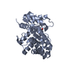
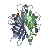
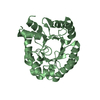
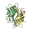
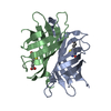
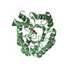
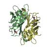
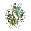
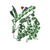
| Title | OBSERVATION OF COVALENT INTERMEDIATES IN AN ENZYME MECHANISM AT ATOMIC RESOLUTION | ||||||
 Components Components | DEOXYRIBOSE-PHOSPHATE ALDOLASE | ||||||
 Keywords Keywords | LYASE / alpha-beta TIM barrel | ||||||
| Function / homology |  Function and homology information Function and homology informationdeoxyribose-phosphate aldolase / deoxyribose-phosphate aldolase activity / 2-deoxyribose 1-phosphate catabolic process / deoxyribonucleotide catabolic process / nucleobase-containing small molecule interconversion / carbohydrate catabolic process / lyase activity / DNA damage response / membrane / cytosol Similarity search - Function | ||||||
| Biological species |  | ||||||
| Method |  X-RAY DIFFRACTION / SYNCHROTRON / FOURIER SYNTHESIS / Resolution: 1.05 Å X-RAY DIFFRACTION / SYNCHROTRON / FOURIER SYNTHESIS / Resolution: 1.05 Å | ||||||
 Authors Authors | Heine, A. / DeSantis, G. / Luz, J.G. / Mitchell, M. / Wong, C.-H. / Wilson, I.A. | ||||||
 Citation Citation | Journal: Science / Year: 2001 Title: Observation of covalent intermediates in an enzyme mechanism at atomic resolution. Authors: Heine, A. / DeSantis, G. / Luz, J.G. / Mitchell, M. / Wong, C.H. / Wilson, I.A. | ||||||
| History |
|
- Structure visualization
| Structure viewer | Molecule: MolmilJmol/JSmol |
|---|
- Downloads & links
-Download
| PDBx/mmCIF format | 1jcl.cif.gz | 230.5 KB | Display | PDBx/mmCIF format |
|---|---|---|---|---|
| PDB format | pdb1jcl.ent.gz | 182.3 KB | Display | PDB format |
| PDBx/mmJSON format | 1jcl.json.gz | Tree view | PDBx/mmJSON format | |
| Others |  Other downloads Other downloads |
-Validation report
| Arichive directory | https://data.pdbj.org/pub/pdb/validation_reports/jc/1jclftp://data.pdbj.org/pub/pdb/validation_reports/jc/1jcl | HTTPS FTP |
|---|
-Related structure data


-Links
 PDBj
PDBj
- Assembly
| Deposited unit | 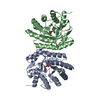
| ||||||||
|---|---|---|---|---|---|---|---|---|---|
| 1 | 
| ||||||||
| 2 | 
| ||||||||
| Unit cell |
|
-Components
| #1: Protein | Mass: 27906.963 Da / Num. of mol.: 2 Source method: isolated from a genetically manipulated source Source: (gene. exp.) #2: Chemical |   Mass: 216.126 Da / Num. of mol.: 2 / Source method: obtained synthetically / Formula: C5H13O7P Mass: 216.126 Da / Num. of mol.: 2 / Source method: obtained synthetically / Formula: C5H13O7P#3: Water | ChemComp-HOH / |  Mass: 18.015 Da / Num. of mol.: 708 / Source method: isolated from a natural source / Formula: H2O Mass: 18.015 Da / Num. of mol.: 708 / Source method: isolated from a natural source / Formula: H2OHas protein modification | Y | |
|---|
-Experimental details
-Experiment
| Experiment | Method: X-RAY DIFFRACTION / Number of used crystals: 1 |
|---|
- Sample preparation
| Crystal | Density Matthews: 2.62 Å3/Da / Density % sol: 52.98 % | |||||||||||||||||||||||||
|---|---|---|---|---|---|---|---|---|---|---|---|---|---|---|---|---|---|---|---|---|---|---|---|---|---|---|
| Crystal grow | Temperature: 277 K / Method: vapor diffusion, sitting drop / pH: 5.5 Details: MPEG 5000, pH 5.5, VAPOR DIFFUSION, SITTING DROP, temperature 277K | |||||||||||||||||||||||||
| Crystal grow | *PLUS Temperature: 4 ℃ / Method: unknown | |||||||||||||||||||||||||
| Components of the solutions | *PLUS
|
-Data collection
| Diffraction | Mean temperature: 97 K |
|---|---|
| Diffraction source | Source: SYNCHROTRON / Site: SSRL  / Beamline: BL9-2 / Wavelength: 1 Å / Beamline: BL9-2 / Wavelength: 1 Å |
| Detector | Type: ADSC QUANTUM 4 / Detector: CCD / Date: Mar 5, 2000 |
| Radiation | Protocol: SINGLE WAVELENGTH / Monochromatic (M) / Laue (L): M / Scattering type: x-ray |
| Radiation wavelength | Wavelength: 1 Å / Relative weight: 1 |
| Reflection | Resolution: 1.05→30 Å / Num. all: 256887 / Num. obs: 256887 / % possible obs: 95.5 % / Redundancy: 2.5 % / Rsym value: 0.063 / Net I/σ(I): 14.8 |
| Reflection shell | Resolution: 1.05→1.07 Å / Mean I/σ(I) obs: 1 / Rsym value: 0.667 / % possible all: 64 |
| Reflection | *PLUS Num. measured all: 638223 / Rmerge(I) obs: 0.063 |
| Reflection shell | *PLUS % possible obs: 64 % / Rmerge(I) obs: 0.667 |
- Processing
| Software |
| |||||||||||||||||||||||||||||||||
|---|---|---|---|---|---|---|---|---|---|---|---|---|---|---|---|---|---|---|---|---|---|---|---|---|---|---|---|---|---|---|---|---|---|---|
| Refinement | Method to determine structure: FOURIER SYNTHESIS Starting model: native DERA Resolution: 1.05→10 Å / Num. parameters: 40900 / Num. restraintsaints: 48367 / Cross valid method: R-free / σ(F): 4 / Stereochemistry target values: ENGH & HUBER
| |||||||||||||||||||||||||||||||||
| Solvent computation | Solvent model: MOEWS & KRETSINGER, J.MOL.BIOL.91(1973)201 | |||||||||||||||||||||||||||||||||
| Refine analyze | Num. disordered residues: 7 | |||||||||||||||||||||||||||||||||
| Refinement step | Cycle: LAST / Resolution: 1.05→10 Å
| |||||||||||||||||||||||||||||||||
| Refine LS restraints |
| |||||||||||||||||||||||||||||||||
| LS refinement shell | Resolution: 1.05→10 Å /
| |||||||||||||||||||||||||||||||||
| Software | *PLUS Name: SHELXL-97 / Classification: refinement | |||||||||||||||||||||||||||||||||
| Refinement | *PLUS Lowest resolution: 10 Å / % reflection Rfree: 5 % / Rfactor all: 0.143 / Rfactor Rwork: 0.143 | |||||||||||||||||||||||||||||||||
| Solvent computation | *PLUS | |||||||||||||||||||||||||||||||||
| Displacement parameters | *PLUS |
