 Movie
Movie Controller

[English] 日本語
 Yorodumi
- PDB-1x8r: EPSPS liganded with the (S)-phosphonate analog of the tetrahedral... -
+ Open data

- Basic information
| Entry | Database: PDB / ID: 1x8r | ||||||
|---|---|---|---|---|---|---|---|
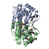
| Title | EPSPS liganded with the (S)-phosphonate analog of the tetrahedral reaction intermediate | ||||||
 Components Components | 3-phosphoshikimate 1-carboxyvinyltransferase | ||||||
 Keywords Keywords | TRANSFERASE / inside-out alpha-beta barrel | ||||||
| Function / homology |  Function and homology information Function and homology information3-phosphoshikimate 1-carboxyvinyltransferase / 3-phosphoshikimate 1-carboxyvinyltransferase activity / chorismate biosynthetic process / aromatic amino acid biosynthetic process / amino acid biosynthetic process / cytosol Similarity search - Function | ||||||
| Biological species |  | ||||||
| Method |  X-RAY DIFFRACTION / MOLECULAR REPLACEMENT / Resolution: 1.5 Å X-RAY DIFFRACTION / MOLECULAR REPLACEMENT / Resolution: 1.5 Å | ||||||
 Authors Authors | Priestman, M.A. / Healy, M.L. / Becker, A. / Alberg, D.G. / Bartlett, P.A. / Schonbrunn, E. | ||||||
 Citation Citation | Journal: Biochemistry / Year: 2005 Title: Interaction of phosphonate analogues of the tetrahedral reaction intermediate with 5-enolpyruvylshikimate-3-phosphate synthase in atomic detail. Authors: Priestman, M.A. / Healy, M.L. / Becker, A. / Alberg, D.G. / Bartlett, P.A. / Lushington, G.H. / Schonbrunn, E. | ||||||
| History |
|
- Structure visualization
| Structure viewer | Molecule: MolmilJmol/JSmol |
|---|
- Downloads & links
-Download
| PDBx/mmCIF format | 1x8r.cif.gz | 110 KB | Display | PDBx/mmCIF format |
|---|---|---|---|---|
| PDB format | pdb1x8r.ent.gz | 82.2 KB | Display | PDB format |
| PDBx/mmJSON format | 1x8r.json.gz | Tree view | PDBx/mmJSON format | |
| Others |  Other downloads Other downloads |
-Validation report
| Arichive directory | https://data.pdbj.org/pub/pdb/validation_reports/x8/1x8rftp://data.pdbj.org/pub/pdb/validation_reports/x8/1x8r | HTTPS FTP |
|---|
-Related structure data
| Related structure data |  1x8tC 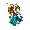 1g6sS S: Starting model for refinement C: citing same article ( |
|---|---|
| Similar structure data |


-Links
 PDBj
PDBj
- Assembly
| Deposited unit | 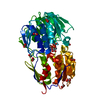
| ||||||||
|---|---|---|---|---|---|---|---|---|---|
| 1 |
| ||||||||
| Unit cell |
|
-Components
| #1: Protein | Mass: 46141.613 Da / Num. of mol.: 1 Source method: isolated from a genetically manipulated source Source: (gene. exp.) References: UniProt: P0A6D3, 3-phosphoshikimate 1-carboxyvinyltransferase | ||
|---|---|---|---|
| #2: Chemical | ChemComp-SC1 / [  Mass: 406.174 Da / Num. of mol.: 1 / Source method: obtained synthetically / Formula: C10H16O13P2 Mass: 406.174 Da / Num. of mol.: 1 / Source method: obtained synthetically / Formula: C10H16O13P2 | ||
| #3: Chemical | ChemComp-FMT /   Mass: 46.025 Da / Num. of mol.: 12 / Source method: obtained synthetically / Formula: CH2O2 Mass: 46.025 Da / Num. of mol.: 12 / Source method: obtained synthetically / Formula: CH2O2#4: Water | ChemComp-HOH / |  Mass: 18.015 Da / Num. of mol.: 568 / Source method: isolated from a natural source / Formula: H2O Mass: 18.015 Da / Num. of mol.: 568 / Source method: isolated from a natural source / Formula: H2O |
-Experimental details
-Experiment
| Experiment | Method: X-RAY DIFFRACTION / Number of used crystals: 1 |
|---|
- Sample preparation
| Crystal | Density Matthews: 2.3 Å3/Da / Density % sol: 46 % |
|---|---|
| Crystal grow | Temperature: 290 K / Method: vapor diffusion, hanging drop / pH: 7 Details: Na-formate, pH 7, VAPOR DIFFUSION, HANGING DROP, temperature 290K |
-Data collection
| Diffraction | Mean temperature: 90 K |
|---|---|
| Diffraction source | Source: ROTATING ANODE / Type: RIGAKU RU300 / Wavelength: 1.5418 Å |
| Detector | Type: RIGAKU RAXIS IV / Detector: IMAGE PLATE / Date: Apr 11, 2004 / Details: mirrors |
| Radiation | Monochromator: osmic mirrors / Protocol: SINGLE WAVELENGTH / Monochromatic (M) / Laue (L): M / Scattering type: x-ray |
| Radiation wavelength | Wavelength: 1.5418 Å / Relative weight: 1 |
| Reflection | Resolution: 1.5→10 Å / Num. all: 66430 / Num. obs: 66430 / % possible obs: 95.3 % / Observed criterion σ(F): 0 / Observed criterion σ(I): -3 / Redundancy: 6.8 % / Rmerge(I) obs: 0.048 / Net I/σ(I): 18.3 |
| Reflection shell | Resolution: 1.5→1.6 Å / Redundancy: 6 % / Rmerge(I) obs: 0.143 / Mean I/σ(I) obs: 7.1 / Num. unique all: 11149 / % possible all: 92.1 |
- Processing
| Software |
| |||||||||||||||||||||||||
|---|---|---|---|---|---|---|---|---|---|---|---|---|---|---|---|---|---|---|---|---|---|---|---|---|---|---|
| Refinement | Method to determine structure: MOLECULAR REPLACEMENT Starting model: PDB entry 1G6S Resolution: 1.5→10 Å / Cross valid method: THROUGHOUT / σ(F): 0 / σ(I): -3 / Stereochemistry target values: Engh & Huber
| |||||||||||||||||||||||||
| Refinement step | Cycle: LAST / Resolution: 1.5→10 Å
| |||||||||||||||||||||||||
| Refine LS restraints |
|
