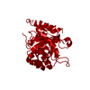
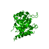
 Movie
Movie Controller


+ Open data

- Basic information
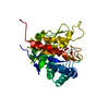
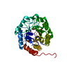
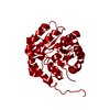
| Entry | Database: PDB / ID: 1x6u | |||||||||
|---|---|---|---|---|---|---|---|---|---|---|
| Title | KDO8P synthase in it's binary complex with the product KDO8P | |||||||||
 Components Components | 2-dehydro-3-deoxyphosphooctonate aldolase | |||||||||
 Keywords Keywords | TRANSFERASE / KDO8P | |||||||||
| Function / homology |  Function and homology information Function and homology information3-deoxy-8-phosphooctulonate synthase / 3-deoxy-8-phosphooctulonate synthase activity / keto-3-deoxy-D-manno-octulosonic acid biosynthetic process / protein-containing complex / identical protein binding / cytosol Similarity search - Function | |||||||||
| Biological species |  | |||||||||
| Method |  X-RAY DIFFRACTION / SYNCHROTRON / MOLECULAR REPLACEMENT / Resolution: 2.7 Å X-RAY DIFFRACTION / SYNCHROTRON / MOLECULAR REPLACEMENT / Resolution: 2.7 Å | |||||||||
 Authors Authors | Vainer, R. / Belakhov, V. / Rabkin, E. / Baasov, T. / Adir, N. | |||||||||
 Citation Citation | Journal: J.Mol.Biol. / Year: 2005 Title: Crystal Structures of Escherichia coli KDO8P Synthase Complexes Reveal the Source of Catalytic Irreversibility Authors: Vainer, R. / Belakhov, V. / Rabkin, E. / Baasov, T. / Adir, N. | |||||||||
| History |
|
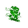
- Structure visualization
| Structure viewer | Molecule: MolmilJmol/JSmol |
|---|
- Downloads & links
-Download
| PDBx/mmCIF format | 1x6u.cif.gz | 65.8 KB | Display | PDBx/mmCIF format |
|---|---|---|---|---|
| PDB format | pdb1x6u.ent.gz | 48.9 KB | Display | PDB format |
| PDBx/mmJSON format | 1x6u.json.gz | Tree view | PDBx/mmJSON format | |
| Others |  Other downloads Other downloads |
-Validation report
| Summary document | 1x6u_validation.pdf.gz | 831.5 KB | Display | wwPDB validaton report |
|---|---|---|---|---|
| Full document | 1x6u_full_validation.pdf.gz | 843 KB | Display | |
| Data in XML | 1x6u_validation.xml.gz | 16 KB | Display | |
| Data in CIF | 1x6u_validation.cif.gz | 19.9 KB | Display | |
| Arichive directory | https://data.pdbj.org/pub/pdb/validation_reports/x6/1x6uftp://data.pdbj.org/pub/pdb/validation_reports/x6/1x6u | HTTPS FTP |
-Related structure data
| Related structure data |  1phwSC 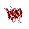 1q3nC  1x8fC S: Starting model for refinement C: citing same article ( |
|---|---|
| Similar structure data |


-Links
 PDBj
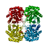
PDBj- Assembly
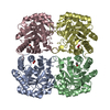
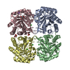
| Deposited unit | 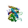
| ||||||||
|---|---|---|---|---|---|---|---|---|---|
| 1 | 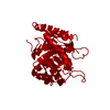
| ||||||||
| Unit cell |
| ||||||||
| Details | The biological assembly is a tetramer generated from the dimer in the asymmetric unit by the operations: X,Y,Z -X,-Y,Z -X,Y,-Z X,-Y,-Z Z,X,Y Z,-X,-Y -Z,-X,Y -Z,X,-Y Y,Z,X -Y,Z,-X Y,-Z,-X -Y,-Z,X 1/2+X,1/2+Y,1/2+Z 1/2-X,1/2-Y,1/2+Z 1/2-X,1/2+Y,1/2-Z 1/2+X,1/2-Y,1/2-Z 1/2+Z,1/2+X,1/2+Y 1/2+Z,1/2-X,1/2-Y 1/2-Z,1/2-X,1/2+Y 1/2-Z,1/2+X,1/2-Y 1/2+Y,1/2+Z,1/2+X 1/2-Y,1/2+Z,1/2-X 1/2+Y,1/2-Z,1/2-X 1/2-Y,1/2-Z,1/2+X |
-Components
| #1: Protein | Mass: 30870.676 Da / Num. of mol.: 1 Source method: isolated from a genetically manipulated source Source: (gene. exp.) References: UniProt: P0A715, 3-deoxy-8-phosphooctulonate synthase |
|---|---|
| #2: Sugar | ChemComp-DO8 / 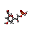  Type: D-saccharide, alpha linking / Mass: 318.172 Da / Num. of mol.: 1 / Source method: obtained synthetically / Formula: C8H15O11P Type: D-saccharide, alpha linking / Mass: 318.172 Da / Num. of mol.: 1 / Source method: obtained synthetically / Formula: C8H15O11P |
| #3: Water | ChemComp-HOH /  Mass: 18.015 Da / Num. of mol.: 25 / Source method: isolated from a natural source / Formula: H2O Mass: 18.015 Da / Num. of mol.: 25 / Source method: isolated from a natural source / Formula: H2O |
| Has protein modification | N |
-Experimental details
-Experiment
| Experiment | Method: X-RAY DIFFRACTION / Number of used crystals: 1 |
|---|
- Sample preparation
| Crystal | Density Matthews: 2.2 Å3/Da / Density % sol: 42.8 % |
|---|---|
| Crystal grow | Temperature: 293 K / Method: vapor diffusion, hanging drop / pH: 7.4 Details: PEG4000, Glycerol, Tris-HCl, pH 7.4, VAPOR DIFFUSION, HANGING DROP, temperature 293K |
-Data collection
| Diffraction | Mean temperature: 100 K |
|---|---|
| Diffraction source | Source: SYNCHROTRON / Site: ESRF  / Beamline: BM30A / Wavelength: 0.934 Å / Beamline: BM30A / Wavelength: 0.934 Å |
| Detector | Type: MARRESEARCH / Detector: CCD / Date: Nov 28, 2003 |
| Radiation | Protocol: SINGLE WAVELENGTH / Monochromatic (M) / Laue (L): M / Scattering type: x-ray |
| Radiation wavelength | Wavelength: 0.934 Å / Relative weight: 1 |
| Reflection | Resolution: 2.7→20 Å / Num. obs: 6950 / % possible obs: 90.7 % / Observed criterion σ(F): 1 / Observed criterion σ(I): 1 / Redundancy: 6.2 % / Rsym value: 0.075 / Net I/σ(I): 13.1 |
| Reflection shell | Resolution: 2.7→2.8 Å / Redundancy: 7 % / Rmerge(I) obs: 0.307 / Mean I/σ(I) obs: 5.3 / Num. unique all: 768 / % possible all: 99.9 |
- Processing
| Software |
| |||||||||||||||||||||||||
|---|---|---|---|---|---|---|---|---|---|---|---|---|---|---|---|---|---|---|---|---|---|---|---|---|---|---|
| Refinement | Method to determine structure: MOLECULAR REPLACEMENT Starting model: PDB ENTRY 1PHW Resolution: 2.7→20 Å / Isotropic thermal model: Isotropic / Cross valid method: THROUGHOUT / σ(F): 3 / Stereochemistry target values: Engh & Huber
| |||||||||||||||||||||||||
| Displacement parameters | Biso mean: 66.35 Å2 | |||||||||||||||||||||||||
| Refine analyze |
| |||||||||||||||||||||||||
| Refinement step | Cycle: LAST / Resolution: 2.7→20 Å
| |||||||||||||||||||||||||
| Refine LS restraints |
|
