 Movie
Movie Controller


+ Open data

- Basic information
| Entry | Database: PDB / ID: 1gg0 | ||||||
|---|---|---|---|---|---|---|---|
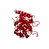
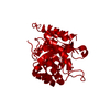
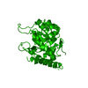
| Title | CRYSTAL STRUCTURE ANALYSIS OF KDOP SYNTHASE AT 3.0 A | ||||||
 Components Components | 3-DEOXY-D-MANNO-OCTULOSONATE 8-PHOSPHATE SYNTHASE | ||||||
 Keywords Keywords | LYASE / beta-alpha-barrel | ||||||
| Function / homology |  Function and homology information Function and homology information3-deoxy-8-phosphooctulonate synthase / 3-deoxy-8-phosphooctulonate synthase activity / keto-3-deoxy-D-manno-octulosonic acid biosynthetic process / protein-containing complex / identical protein binding / cytosol Similarity search - Function | ||||||
| Biological species |  | ||||||
| Method |  X-RAY DIFFRACTION / Resolution: 3 Å X-RAY DIFFRACTION / Resolution: 3 Å | ||||||
 Authors Authors | Wagner, T. / Kretsinger, R.H. / Bauerle, R. / Tolbert, W.D. | ||||||
 Citation Citation | Journal: J.Mol.Biol. / Year: 2000 Title: 3-Deoxy-D-manno-octulosonate-8-phosphate synthase from Escherichia coli. Model of binding of phosphoenolpyruvate and D-arabinose-5-phosphate. Authors: Wagner, T. / Kretsinger, R.H. / Bauerle, R. / Tolbert, W.D. #1: Journal: J.Biol.Chem. / Year: 2000Title: Structure and mechanism of 3-deoxy-D-manno-octulosonate-8-phosphate synthase Authors: Radaev, S. / Dastidar, P. / Patel, M. / Woodard, R.W. / Gatti, D.L. #2: Journal: Proteins / Year: 1996Title: Crystallization and preliminary crystallographic studies of 3-deoxy-D-manno-octulosonate-8-phosphate synthase from Escherichia coli Authors: Tolbert, W.D. / Moll, J.R. / Bauerle, R. / Kretsinger, R.H. | ||||||
| History |
|
- Structure visualization
| Structure viewer | Molecule: MolmilJmol/JSmol |
|---|
- Downloads & links
-Download
| PDBx/mmCIF format | 1gg0.cif.gz | 58.3 KB | Display | PDBx/mmCIF format |
|---|---|---|---|---|
| PDB format | pdb1gg0.ent.gz | 44.3 KB | Display | PDB format |
| PDBx/mmJSON format | 1gg0.json.gz | Tree view | PDBx/mmJSON format | |
| Others |  Other downloads Other downloads |
-Validation report
| Arichive directory | https://data.pdbj.org/pub/pdb/validation_reports/gg/1gg0ftp://data.pdbj.org/pub/pdb/validation_reports/gg/1gg0 | HTTPS FTP |
|---|
-Related structure data
| Related structure data | |
|---|---|
| Similar structure data |


-Links
 PDBj
PDBj- Assembly
| Deposited unit | 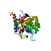
| ||||||||
|---|---|---|---|---|---|---|---|---|---|
| 1 | 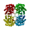
| ||||||||
| 2 | 
| ||||||||
| Unit cell |
| ||||||||
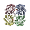
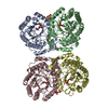
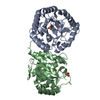
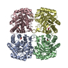
| Details | The biological assembly is a tetramer constructed from chain A and the symmetry partners generated by two two-fold axes. |
-Components
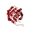
| #1: Protein | Mass: 30870.676 Da / Num. of mol.: 1 Source method: isolated from a genetically manipulated source Source: (gene. exp.) |
|---|---|
| #2: Chemical |   Mass: 94.971 Da / Num. of mol.: 2 / Source method: obtained synthetically / Formula: PO4 Mass: 94.971 Da / Num. of mol.: 2 / Source method: obtained synthetically / Formula: PO4 |
-Experimental details
-Experiment
| Experiment | Method: X-RAY DIFFRACTION / Number of used crystals: 1 |
|---|
- Sample preparation
| Crystal | Density Matthews: 2.23 Å3/Da / Density % sol: 44.74 % | ||||||||||||||||||||
|---|---|---|---|---|---|---|---|---|---|---|---|---|---|---|---|---|---|---|---|---|---|
| Crystal grow | Temperature: 291 K / Method: vapor diffusion, hanging drop / pH: 7 Details: PEG 1500, 3-(N-morpholino)propanesulfonic acid, dithioerythitol, d-arabinose-5-phosphate, pH 7.0, VAPOR DIFFUSION, HANGING DROP, temperature 291.0K | ||||||||||||||||||||
| Crystal grow | *PLUS Details: drop consists of equal amounts of protein and reservoir solutions | ||||||||||||||||||||
| Components of the solutions | *PLUS
|
-Data collection
| Diffraction | Mean temperature: 277 K |
|---|---|
| Diffraction source | Source: ROTATING ANODE / Type: OTHER / Wavelength: 1.5418 |
| Detector | Type: CUSTOM-MADE / Detector: AREA DETECTOR / Date: Mar 13, 1996 |
| Radiation | Protocol: SINGLE WAVELENGTH / Monochromatic (M) / Laue (L): M / Scattering type: x-ray |
| Radiation wavelength | Wavelength: 1.5418 Å / Relative weight: 1 |
| Reflection | Resolution: 3→20 Å / Num. obs: 4988 / % possible obs: 88.6 % / Observed criterion σ(I): 2 / Redundancy: 7.8 % / Biso Wilson estimate: 52.2 Å2 / Rmerge(I) obs: 0.054 / Net I/σ(I): 43.8 |
| Reflection shell | Resolution: 3→3.11 Å / Redundancy: 3.4 % / Rmerge(I) obs: 0.116 / Mean I/σ(I) obs: 9.8 / Num. unique all: 357 / % possible all: 62.9 |
| Reflection | *PLUS Num. measured all: 39075 |
| Reflection shell | *PLUS % possible obs: 62.9 % |
- Processing
| Software |
| ||||||||||||||||||||
|---|---|---|---|---|---|---|---|---|---|---|---|---|---|---|---|---|---|---|---|---|---|
| Refinement | Resolution: 3→20 Å / σ(I): 2 / Stereochemistry target values: Engh & Huber
| ||||||||||||||||||||
| Refinement step | Cycle: LAST / Resolution: 3→20 Å
| ||||||||||||||||||||
| Refine LS restraints |
| ||||||||||||||||||||
| Software | *PLUS Name: CNS / Classification: refinement | ||||||||||||||||||||
| Refinement | *PLUS % reflection Rfree: 9 % | ||||||||||||||||||||
| Solvent computation | *PLUS | ||||||||||||||||||||
| Displacement parameters | *PLUS |
