 Movie
Movie Controller


+ Open data

- Basic information
| Entry | Database: PDB / ID: 1x3w | |||||||||
|---|---|---|---|---|---|---|---|---|---|---|
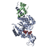
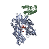
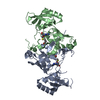
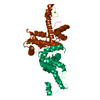
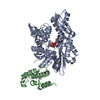
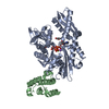
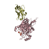
| Title | Structure of a peptide:N-glycanase-Rad23 complex | |||||||||
 Components Components |
| |||||||||
 Keywords Keywords | HYDROLASE / Protein-protein complex | |||||||||
| Function / homology |  Function and homology information Function and homology informationubiquitin-dependent glycoprotein ERAD pathway / peptide-N4-(N-acetyl-beta-glucosaminyl)asparagine amidase / peptide-N4-(N-acetyl-beta-glucosaminyl)asparagine amidase activity / nucleotide-excision repair factor 2 complex / nucleotide-excision repair, DNA damage recognition / protein deglycosylation / K48-linked polyubiquitin modification-dependent protein binding / proteasome binding / protein quality control for misfolded or incompletely synthesized proteins / polyubiquitin modification-dependent protein binding ...ubiquitin-dependent glycoprotein ERAD pathway / peptide-N4-(N-acetyl-beta-glucosaminyl)asparagine amidase / peptide-N4-(N-acetyl-beta-glucosaminyl)asparagine amidase activity / nucleotide-excision repair factor 2 complex / nucleotide-excision repair, DNA damage recognition / protein deglycosylation / K48-linked polyubiquitin modification-dependent protein binding / proteasome binding / protein quality control for misfolded or incompletely synthesized proteins / polyubiquitin modification-dependent protein binding / ERAD pathway / ubiquitin binding / damaged DNA binding / proteasome-mediated ubiquitin-dependent protein catabolic process / protein-macromolecule adaptor activity / negative regulation of transcription by RNA polymerase II / mitochondrion / nucleoplasm / metal ion binding / nucleus / cytosol / cytoplasm Similarity search - Function | |||||||||
| Biological species |  | |||||||||
| Method |  X-RAY DIFFRACTION / SYNCHROTRON / SAD / Resolution: 3 Å X-RAY DIFFRACTION / SYNCHROTRON / SAD / Resolution: 3 Å | |||||||||
 Authors Authors | Lee, J.-H. / Choi, J.M. / Lee, C. / Yi, K.J. / Cho, Y. | |||||||||
 Citation Citation | Journal: Proc.Natl.Acad.Sci.Usa / Year: 2005 Title: Structure of a peptide:N-glycanase-Rad23 complex: insight into the deglycosylation for denatured glycoproteins. Authors: Lee, J.H. / Choi, J.M. / Lee, C. / Yi, K.J. / Cho, Y. | |||||||||
| History |
|
- Structure visualization
| Structure viewer | Molecule: MolmilJmol/JSmol |
|---|
- Downloads & links
-Download
| PDBx/mmCIF format | 1x3w.cif.gz | 165.4 KB | Display | PDBx/mmCIF format |
|---|---|---|---|---|
| PDB format | pdb1x3w.ent.gz | 130.4 KB | Display | PDB format |
| PDBx/mmJSON format | 1x3w.json.gz | Tree view | PDBx/mmJSON format | |
| Others |  Other downloads Other downloads |
-Validation report
| Arichive directory | https://data.pdbj.org/pub/pdb/validation_reports/x3/1x3wftp://data.pdbj.org/pub/pdb/validation_reports/x3/1x3w | HTTPS FTP |
|---|
-Related structure data


-Links
 PDBj
PDBj


- Assembly
| Deposited unit | 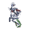
| ||||||||
|---|---|---|---|---|---|---|---|---|---|
| 1 |
| ||||||||
| Unit cell |
| ||||||||
| Details | The biological assembly is protein-protein complex in asymmetric unit |
-Components
| #1: Protein | Mass: 39566.234 Da / Num. of mol.: 1 / Fragment: residues 8-342 Source method: isolated from a genetically manipulated source Source: (gene. exp.) Species (production host): Escherichia coli / Production host:  References: UniProt: Q02890, peptide-N4-(N-acetyl-beta-glucosaminyl)asparagine amidase | ||||||
|---|---|---|---|---|---|---|---|
| #2: Protein | Mass: 7539.189 Da / Num. of mol.: 1 / Fragment: XPC binding domain Source method: isolated from a genetically manipulated source Source: (gene. exp.) Species (production host): Escherichia coli / Production host: | ||||||
| #3: Polysaccharide |   Source method: isolated from a genetically manipulated source Details: oligosaccharide with reducing-end-to-reducing-end glycosidic bond References: sucrose #4: Chemical | ChemComp-ZN / |   Mass: 65.409 Da / Num. of mol.: 1 / Source method: obtained synthetically / Formula: Zn Mass: 65.409 Da / Num. of mol.: 1 / Source method: obtained synthetically / Formula: Zn#5: Water | ChemComp-HOH / |  Mass: 18.015 Da / Num. of mol.: 17 / Source method: isolated from a natural source / Formula: H2O Mass: 18.015 Da / Num. of mol.: 17 / Source method: isolated from a natural source / Formula: H2OHas protein modification | Y | |
-Experimental details
-Experiment
| Experiment | Method: X-RAY DIFFRACTION / Number of used crystals: 1 |
|---|
- Sample preparation
| Crystal | Density Matthews: 6.7 Å3/Da / Density % sol: 81 % |
|---|---|
| Crystal grow | Temperature: 291 K / Method: vapor diffusion / pH: 6 Details: sodium chloride, MES, pH 6, VAPOR DIFFUSION, temperature 291K |
-Data collection
| Diffraction | Mean temperature: 93 K |
|---|---|
| Diffraction source | Source: SYNCHROTRON / Site: Photon Factory  / Beamline: BL-5A / Wavelength: 0.9794 Å / Beamline: BL-5A / Wavelength: 0.9794 Å |
| Radiation | Monochromator: mirror / Protocol: SINGLE WAVELENGTH / Monochromatic (M) / Laue (L): M / Scattering type: x-ray |
| Radiation wavelength | Wavelength: 0.9794 Å / Relative weight: 1 |
| Reflection | Resolution: 3→50 Å / Num. all: 25391 / Num. obs: 25391 / % possible obs: 94.4 % / Observed criterion σ(F): 1 / Observed criterion σ(I): 1 |
| Reflection shell | Resolution: 3→3.1 Å / Rmerge(I) obs: 0.32 / % possible all: 68.9 |
- Processing
| Software |
| |||||||||||||||||||||||||||||||||||||||||||||||||||||||||||||||||||||||||||||||||||||||||||||||||||||||||
|---|---|---|---|---|---|---|---|---|---|---|---|---|---|---|---|---|---|---|---|---|---|---|---|---|---|---|---|---|---|---|---|---|---|---|---|---|---|---|---|---|---|---|---|---|---|---|---|---|---|---|---|---|---|---|---|---|---|---|---|---|---|---|---|---|---|---|---|---|---|---|---|---|---|---|---|---|---|---|---|---|---|---|---|---|---|---|---|---|---|---|---|---|---|---|---|---|---|---|---|---|---|---|---|---|---|---|
| Refinement | Method to determine structure: SAD / Resolution: 3→20 Å / Cor.coef. Fo:Fc: 0.921 / Cor.coef. Fo:Fc free: 0.9 / SU B: 37.386 / SU ML: 0.265 / Cross valid method: THROUGHOUT / σ(F): 0 / ESU R Free: 0.319 / Stereochemistry target values: MAXIMUM LIKELIHOOD / Details: HYDROGENS HAVE BEEN ADDED IN THE RIDING POSITIONS
| |||||||||||||||||||||||||||||||||||||||||||||||||||||||||||||||||||||||||||||||||||||||||||||||||||||||||
| Solvent computation | Ion probe radii: 0.8 Å / Shrinkage radii: 0.8 Å / VDW probe radii: 1.2 Å / Solvent model: MASK | |||||||||||||||||||||||||||||||||||||||||||||||||||||||||||||||||||||||||||||||||||||||||||||||||||||||||
| Displacement parameters | Biso mean: 75.596 Å2
| |||||||||||||||||||||||||||||||||||||||||||||||||||||||||||||||||||||||||||||||||||||||||||||||||||||||||
| Refinement step | Cycle: LAST / Resolution: 3→20 Å
| |||||||||||||||||||||||||||||||||||||||||||||||||||||||||||||||||||||||||||||||||||||||||||||||||||||||||
| Refine LS restraints |
| |||||||||||||||||||||||||||||||||||||||||||||||||||||||||||||||||||||||||||||||||||||||||||||||||||||||||
| LS refinement shell | Resolution: 3→3.076 Å / Total num. of bins used: 20
|
