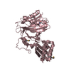
lipoyltransferase activity / lipoate-protein ligase / lipoate-protein ligase activity / protein lipoylation / ATP binding / cytoplasm / cytosol Similarity search - Function
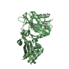
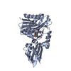
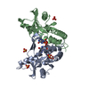
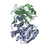
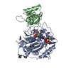
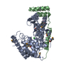
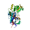
Lipoate-protein ligase A / Lipoate protein ligase, C-terminal / Bacterial lipoate protein ligase C-terminus / Lipoyltransferase/lipoate-protein ligase / Lipoyl protein ligase A/B catalytic domain / CO dehydrogenase flavoprotein, C-terminal domain / Biotinyl protein ligase (BPL) and lipoyl protein ligase (LPL) catalytic domain profile. / Biotin/lipoate A/B protein ligase family / Biotinyl protein ligase (BPL) and lipoyl protein ligase (LPL), catalytic domain / Bira Bifunctional Protein; Domain 2 ...Lipoate-protein ligase A / Lipoate protein ligase, C-terminal / Bacterial lipoate protein ligase C-terminus / Lipoyltransferase/lipoate-protein ligase / Lipoyl protein ligase A/B catalytic domain / CO dehydrogenase flavoprotein, C-terminal domain / Biotinyl protein ligase (BPL) and lipoyl protein ligase (LPL) catalytic domain profile. / Biotin/lipoate A/B protein ligase family / Biotinyl protein ligase (BPL) and lipoyl protein ligase (LPL), catalytic domain / Bira Bifunctional Protein; Domain 2 / BirA Bifunctional Protein; domain 2 / Enolase-like; domain 1 / Class II Aminoacyl-tRNA synthetase/Biotinyl protein ligase (BPL) and lipoyl protein ligase (LPL) / 2-Layer Sandwich / Alpha Beta Similarity search - Domain/homology
Resolution: 2.9→3 Å / Rmerge(I) obs: 0.421 / Mean I/σ(I) obs: 3.8 / Num. unique all: 2939 / % possible all: 99
-
Processing
Software
Name
Version
Classification
REFMAC
5.2.0005
refinement
HKL-2000
datareduction
SCALEPACK
datascaling
Refinement
Method to determine structure: Difference Fourier Starting model: Lipoate protein ligase A from E. coli Resolution: 2.91→66.67 Å / Cor.coef. Fo:Fc: 0.929 / Cor.coef. Fo:Fc free: 0.859 / SU B: 15.864 / SU ML: 0.303 / Cross valid method: THROUGHOUT / σ(F): 0 / ESU R Free: 0.435 / Stereochemistry target values: MAXIMUM LIKELIHOOD
Rfactor
Num. reflection
% reflection
Selection details
Rfree
0.2714
1533
5.3 %
RANDOM
Rwork
0.1863
-
-
-
all
0.1907
27666
-
-
obs
0.1907
27666
97.1 %
-
Solvent computation
Ion probe radii: 0.8 Å / Shrinkage radii: 0.8 Å / VDW probe radii: 1.2 Å / Solvent model: MASK
Displacement parameters
Biso mean: 39.469 Å2
Baniso -1
Baniso -2
Baniso -3
1-
0.59 Å2
0 Å2
0 Å2
2-
-
-0.05 Å2
0 Å2
3-
-
-
-0.54 Å2
Refine analyze
Free
Obs
Luzzati coordinate error
0.303 Å
0.435 Å
Refinement step
Cycle: LAST / Resolution: 2.91→66.67 Å
Protein
Nucleic acid
Ligand
Solvent
Total
Num. atoms
7891
0
24
22
7937
Refine LS restraints
Refine-ID
Type
Dev ideal
Dev ideal target
Number
X-RAY DIFFRACTION
r_bond_refined_d
0.02
0.022
8084
X-RAY DIFFRACTION
r_bond_other_d
X-RAY DIFFRACTION
r_angle_refined_deg
1.976
1.948
10951
X-RAY DIFFRACTION
r_angle_other_deg
X-RAY DIFFRACTION
r_dihedral_angle_1_deg
8.038
5
992
X-RAY DIFFRACTION
r_dihedral_angle_2_deg
35.855
23.596
406
X-RAY DIFFRACTION
r_dihedral_angle_3_deg
21.67
15
1344
X-RAY DIFFRACTION
r_dihedral_angle_4_deg
19.997
15
73
X-RAY DIFFRACTION
r_chiral_restr
0.119
0.2
1196
X-RAY DIFFRACTION
r_gen_planes_refined
0.006
0.02
6240
X-RAY DIFFRACTION
r_gen_planes_other
X-RAY DIFFRACTION
r_nbd_refined
0.254
0.2
3992
X-RAY DIFFRACTION
r_nbd_other
X-RAY DIFFRACTION
r_nbtor_refined
0.329
0.2
5509
X-RAY DIFFRACTION
r_nbtor_other
X-RAY DIFFRACTION
r_xyhbond_nbd_refined
0.169
0.2
329
X-RAY DIFFRACTION
r_xyhbond_nbd_other
X-RAY DIFFRACTION
r_metal_ion_refined
X-RAY DIFFRACTION
r_metal_ion_other
X-RAY DIFFRACTION
r_symmetry_vdw_refined
0.195
0.2
76
X-RAY DIFFRACTION
r_symmetry_vdw_other
X-RAY DIFFRACTION
r_symmetry_hbond_refined
0.169
0.2
7
X-RAY DIFFRACTION
r_symmetry_hbond_other
X-RAY DIFFRACTION
r_symmetry_metal_ion_refined
X-RAY DIFFRACTION
r_symmetry_metal_ion_other
X-RAY DIFFRACTION
r_mcbond_it
0.845
1.5
5048
X-RAY DIFFRACTION
r_mcbond_other
X-RAY DIFFRACTION
r_mcangle_it
1.467
2
7946
X-RAY DIFFRACTION
r_scbond_it
2.238
3
3394
X-RAY DIFFRACTION
r_scangle_it
3.692
4.5
3005
X-RAY DIFFRACTION
r_rigid_bond_restr
X-RAY DIFFRACTION
r_sphericity_free
X-RAY DIFFRACTION
r_sphericity_bonded
LS refinement shell
Highest resolution: 2.91 Å / Num. reflection Rwork: 1955 / Total num. of bins used: 20
+
About Yorodumi
-
News
-
Feb 9, 2022. New format data for meta-information of EMDB entries
New format data for meta-information of EMDB entries
Version 3 of the EMDB header file is now the official format.
The previous official version 1.9 will be removed from the archive.
In the structure databanks used in Yorodumi, some data are registered as the other names, "COVID-19 virus" and "2019-nCoV". Here are the details of the virus and the list of structure data.
Jan 31, 2019. EMDB accession codes are about to change! (news from PDBe EMDB page)
EMDB accession codes are about to change! (news from PDBe EMDB page)
The allocation of 4 digits for EMDB accession codes will soon come to an end. Whilst these codes will remain in use, new EMDB accession codes will include an additional digit and will expand incrementally as the available range of codes is exhausted. The current 4-digit format prefixed with “EMD-” (i.e. EMD-XXXX) will advance to a 5-digit format (i.e. EMD-XXXXX), and so on. It is currently estimated that the 4-digit codes will be depleted around Spring 2019, at which point the 5-digit format will come into force.
The EM Navigator/Yorodumi systems omit the EMD- prefix.
Related info.:Q: What is EMD? / ID/Accession-code notation in Yorodumi/EM Navigator
Yorodumi is a browser for structure data from EMDB, PDB, SASBDB, etc.
This page is also the successor to EM Navigator detail page, and also detail information page/front-end page for Omokage search.
The word "yorodu" (or yorozu) is an old Japanese word meaning "ten thousand". "mi" (miru) is to see.
Related info.:EMDB / PDB / SASBDB / Comparison of 3 databanks / Yorodumi Search / Aug 31, 2016. New EM Navigator & Yorodumi / Yorodumi Papers / Jmol/JSmol / Function and homology information / Changes in new EM Navigator and Yorodumi
 Movie
Movie Controller

 Yorodumi
 Open data
 Basic information
 Components
 Keywords
 Function and homology information
 X-RAY DIFFRACTION /
  Authors
 Citation
 Structure visualization
 Downloads & links
 Other downloads
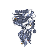


 PDBj
PDBj
 Assembly
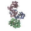


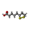
 Mass: 206.326 Da / Num. of mol.: 2 / Source method: obtained synthetically / Formula: C8H14O2S2
 Mass: 18.015 Da / Num. of mol.: 22 / Source method: isolated from a natural source / Formula: H2O
 Sample preparation
 / Beamline: BL44XU / Wavelength: 0.92154 Å
 Processing