[English] 日本語
Yorodumi
- PDB-4tvy: Apo resorufin ligase -

+
Open data


ID or keywords:

Loading...

-
Basic information

Entry
Database: PDB / ID: 4tvy
TitleApo resorufin ligase
ComponentsLipoate-protein ligase ALipoate–protein ligase
Keywords
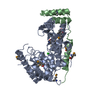
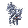
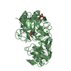
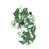
TRANSFERASE / E. coli LplA / computational enzyme design
Function / homology
Function and homology information


lipoyltransferase activity / lipoate-protein ligase / lipoate-protein ligase activity / protein lipoylation / ATP binding / cytosol / cytoplasm
Similarity search - Function
Lipoate-protein ligase A / Lipoate protein ligase, C-terminal / Bacterial lipoate protein ligase C-terminus / Lipoyltransferase/lipoate-protein ligase / CO dehydrogenase flavoprotein, C-terminal domain / Biotinyl protein ligase (BPL) and lipoyl protein ligase (LPL) catalytic domain profile. / Biotin/lipoate A/B protein ligase family / Biotinyl protein ligase (BPL) and lipoyl protein ligase (LPL), catalytic domain / Bira Bifunctional Protein; Domain 2 / BirA Bifunctional Protein; domain 2 ...Lipoate-protein ligase A / Lipoate protein ligase, C-terminal / Bacterial lipoate protein ligase C-terminus / Lipoyltransferase/lipoate-protein ligase / CO dehydrogenase flavoprotein, C-terminal domain / Biotinyl protein ligase (BPL) and lipoyl protein ligase (LPL) catalytic domain profile. / Biotin/lipoate A/B protein ligase family / Biotinyl protein ligase (BPL) and lipoyl protein ligase (LPL), catalytic domain / Bira Bifunctional Protein; Domain 2 / BirA Bifunctional Protein; domain 2 / Class II Aminoacyl-tRNA synthetase/Biotinyl protein ligase (BPL) and lipoyl protein ligase (LPL) / Enolase-like; domain 1 / 2-Layer Sandwich / Alpha Beta
Similarity search - Domain/homology
5-(3,7-dihydroxy-10H-phenoxazin-2-yl)pentanamide / Lipoate-protein ligase A
Similarity search - Component
Biological speciesEscherichia coli (E. coli)
MethodX-RAY DIFFRACTION / SYNCHROTRON / MOLECULAR REPLACEMENT / Resolution: 2.151 Å
AuthorsGoldman, P.J. / Drennan, C.L.
Funding support United States, 1items
OrganizationGrant numberCountry
Howard Hughes Medical Institute (HHMI) United States
CitationJournal: Proc.Natl.Acad.Sci.USA / Year: 2014
Title: Computational design of a red fluorophore ligase for site-specific protein labeling in living cells.
Authors: Liu, D.S. / Nivon, L.G. / Richter, F. / Goldman, P.J. / Deerinck, T.J. / Yao, J.Z. / Richardson, D. / Phipps, W.S. / Ye, A.Z. / Ellisman, M.H. / Drennan, C.L. / Baker, D. / Ting, A.Y.
History
DepositionJun 28, 2014Deposition site: RCSB / Processing site: RCSB
Revision 1.0Oct 22, 2014Provider: repository / Type: Initial release
Revision 1.1Nov 5, 2014Group: Database references
Revision 1.2Sep 13, 2017Group: Author supporting evidence / Database references ...Author supporting evidence / Database references / Derived calculations / Other / Refinement description / Source and taxonomy
Category: citation / entity_src_gen ...citation / entity_src_gen / pdbx_audit_support / pdbx_database_status / pdbx_struct_oper_list / software
Item: _citation.journal_id_CSD / _entity_src_gen.pdbx_alt_source_flag ..._citation.journal_id_CSD / _entity_src_gen.pdbx_alt_source_flag / _pdbx_audit_support.funding_organization / _pdbx_database_status.pdb_format_compatible / _pdbx_struct_oper_list.symmetry_operation / _software.name
Revision 1.3Nov 22, 2017Group: Refinement description / Category: software / Item: _software.classification
Revision 1.4Nov 20, 2019Group: Author supporting evidence / Category: pdbx_audit_support / Item: _pdbx_audit_support.funding_organization
Revision 1.5Sep 27, 2023Group: Data collection / Database references / Refinement description
Category: chem_comp_atom / chem_comp_bond ...chem_comp_atom / chem_comp_bond / database_2 / pdbx_initial_refinement_model / refine_hist
Item: _database_2.pdbx_DOI / _database_2.pdbx_database_accession ..._database_2.pdbx_DOI / _database_2.pdbx_database_accession / _refine_hist.number_atoms_total / _refine_hist.pdbx_number_atoms_nucleic_acid / _refine_hist.pdbx_number_atoms_protein

-
Structure visualization

Structure viewerMolecule:
MolmilJmol/JSmol

Downloads & links

-
Assembly

Deposited unit
A: Lipoate-protein ligase A
B: Lipoate-protein ligase A
hetero molecules


Theoretical massNumber of molelcules
Total (without water)77,3356
Polymers76,0782
Non-polymers1,2574
Water2,900161
1
A: Lipoate-protein ligase A
hetero molecules


Theoretical massNumber of molelcules
Total (without water)38,6683
Polymers38,0391
Non-polymers6292
Water181
TypeNameSymmetry operationNumber
identity operation1_555x,y,z1
2
B: Lipoate-protein ligase A
hetero molecules


Theoretical massNumber of molelcules
Total (without water)38,6683
Polymers38,0391
Non-polymers6292
Water181
TypeNameSymmetry operationNumber
identity operation1_555x,y,z1
Unit cell
Length a, b, c (Å)79.117, 89.951, 108.890
Angle α, β, γ (deg.)90.000, 90.000, 90.000
Int Tables number19
Space group name H-MP212121

-
Components

#1: Protein Lipoate-protein ligase A / Lipoate–protein ligase / Lipoate--protein ligase


Mass: 38038.867 Da / Num. of mol.: 2 / Mutation: E20A, F147, H149G
Source method: isolated from a genetically manipulated source
Source: (gene. exp.) Escherichia coli (E. coli) / Strain: K12 / Gene: lplA, yjjF, b4386, JW4349 / Production host: Escherichia coli (E. coli) / References: UniProt: P32099, EC: 2.7.7.63
#2: Chemical
ChemComp-37R / 5-(3,7-dihydroxy-10H-phenoxazin-2-yl)pentanamide


Mass: 314.336 Da / Num. of mol.: 4 / Source method: obtained synthetically / Formula: C17H18N2O4
#3: Water ChemComp-HOH / water / Water


Mass: 18.015 Da / Num. of mol.: 161 / Source method: isolated from a natural source / Formula: H2O

-
Experimental details

-
Experiment

ExperimentMethod: X-RAY DIFFRACTION

-
Sample preparation

CrystalDensity Matthews: 2.55 Å3/Da / Density % sol: 51.72 %
Crystal growTemperature: 277 K / Method: vapor diffusion, hanging drop
Details: 2 uL of 5.6 mg/mL resorufin ligase containing 1 mM resorufin sulfamoyladenosine, 1 mM Mg(OAc)2, and 1 mM dithiothreitol mixed with 2 uL of precipitant solution (11% PEG 20,000, 0.15 M MES: ...Details: 2 uL of 5.6 mg/mL resorufin ligase containing 1 mM resorufin sulfamoyladenosine, 1 mM Mg(OAc)2, and 1 mM dithiothreitol mixed with 2 uL of precipitant solution (11% PEG 20,000, 0.15 M MES:NaOH, pH 6.5). Red colored crystalline plates appeared after ~ 5 days. Crystals were looped and washed through a cryoprotection solution of 80% precipitant solution (12% PEG 20,000, 0.2 M MES:NaOH, pH 6.5) and 20% glycerol. Crystals were then cryocooled by direction submersion into liquid nitrogen.

-
Data collection

DiffractionMean temperature: 100 K
Diffraction sourceSource: SYNCHROTRON / Site: APS / Beamline: 24-ID-C / Wavelength: 0.9795 Å
DetectorType: DECTRIS PILATUS 6M / Detector: PIXEL / Date: Oct 15, 2012
RadiationProtocol: SINGLE WAVELENGTH / Monochromatic (M) / Laue (L): M / Scattering type: x-ray
Radiation wavelengthWavelength: 0.9795 Å / Relative weight: 1
ReflectionResolution: 2.15→44.98 Å / Num. obs: 42531 / % possible obs: 99.1 % / Redundancy: 5.4 % / Biso Wilson estimate: 38.63 Å2 / Rmerge(I) obs: 0.067 / Net I/σ(I): 13
Reflection shellResolution: 2.15→2.23 Å / Redundancy: 4.6 % / Mean I/σ(I) obs: 2.25 / Rsym value: 0.541 / % possible all: 95

-
Processing

Software
NameVersionClassification
PHENIX(phenix.refine: 1.9_1692)refinement
PHASERphasing
RefinementMethod to determine structure: MOLECULAR REPLACEMENT
Starting model: 1X2G
Resolution: 2.151→44.976 Å / SU ML: 0.23 / Cross valid method: NONE / σ(F): 1.34 / Phase error: 28.65 / Stereochemistry target values: ML
RfactorNum. reflection% reflection
Rfree0.2615 2144 5.05 %
Rwork0.212 40353 -
obs0.2145 42497 99 %
Solvent computationShrinkage radii: 0.9 Å / VDW probe radii: 1.11 Å / Solvent model: FLAT BULK SOLVENT MODEL
Displacement parametersBiso max: 94.9 Å2 / Biso mean: 45.3337 Å2 / Biso min: 24.04 Å2
Refinement stepCycle: final / Resolution: 2.151→44.976 Å
ProteinNucleic acidLigandSolventTotal
Num. atoms5118 0 138 161 5417
Biso mean--56.53 42.89 -
Num. residues----650
Refine LS restraints
Refine-IDTypeDev idealNumber
X-RAY DIFFRACTIONf_bond_d0.0065335
X-RAY DIFFRACTIONf_angle_d1.0337243
X-RAY DIFFRACTIONf_chiral_restr0.062779
X-RAY DIFFRACTIONf_plane_restr0.004948
X-RAY DIFFRACTIONf_dihedral_angle_d15.8551927
LS refinement shell

Refine-ID: X-RAY DIFFRACTION / Total num. of bins used: 15

Resolution (Å)Rfactor RfreeNum. reflection RfreeRfactor RworkNum. reflection RworkNum. reflection all% reflection obs (%)
2.1509-2.20090.35911390.28822518265793
2.2009-2.2560.33261310.26792663279499
2.256-2.3170.30171380.265926842822100
2.317-2.38510.32941500.264526532803100
2.3851-2.46210.31811800.260126322812100
2.4621-2.55010.34281680.25812648281699
2.5501-2.65220.30481420.248527012843100
2.6522-2.77290.29491200.235726952815100
2.7729-2.91910.30111240.244127112835100
2.9191-3.10190.2951320.23672719285199
3.1019-3.34130.26461320.2212701283399
3.3413-3.67740.24941420.203727152857100
3.6774-4.20920.20561460.18722706285299
4.2092-5.30190.23161450.16932777292299
5.3019-44.98530.22961550.18892830298598

+
About Yorodumi

-
News

-
Feb 9, 2022. New format data for meta-information of EMDB entries

New format data for meta-information of EMDB entries

  • Version 3 of the EMDB header file is now the official format.
  • The previous official version 1.9 will be removed from the archive.

Related info.:EMDB header

External links:wwPDB to switch to version 3 of the EMDB data model

-
Aug 12, 2020. Covid-19 info

Covid-19 info

URL: https://pdbj.org/emnavi/covid19.php

New page: Covid-19 featured information page in EM Navigator.

Related info.:Covid-19 info / Mar 5, 2020. Novel coronavirus structure data

+
Mar 5, 2020. Novel coronavirus structure data

Novel coronavirus structure data

Related info.:Yorodumi Speices / Aug 12, 2020. Covid-19 info

External links:COVID-19 featured content - PDBj / Molecule of the Month (242):Coronavirus Proteases

+
Jan 31, 2019. EMDB accession codes are about to change! (news from PDBe EMDB page)

EMDB accession codes are about to change! (news from PDBe EMDB page)

  • The allocation of 4 digits for EMDB accession codes will soon come to an end. Whilst these codes will remain in use, new EMDB accession codes will include an additional digit and will expand incrementally as the available range of codes is exhausted. The current 4-digit format prefixed with “EMD-” (i.e. EMD-XXXX) will advance to a 5-digit format (i.e. EMD-XXXXX), and so on. It is currently estimated that the 4-digit codes will be depleted around Spring 2019, at which point the 5-digit format will come into force.
  • The EM Navigator/Yorodumi systems omit the EMD- prefix.

Related info.:Q: What is EMD? / ID/Accession-code notation in Yorodumi/EM Navigator

External links:EMDB Accession Codes are Changing Soon! / Contact to PDBj

+
Jul 12, 2017. Major update of PDB

Major update of PDB

  • wwPDB released updated PDB data conforming to the new PDBx/mmCIF dictionary.
  • This is a major update changing the version number from 4 to 5, and with Remediation, in which all the entries are updated.
  • In this update, many items about electron microscopy experimental information are reorganized (e.g. em_software).
  • Now, EM Navigator and Yorodumi are based on the updated data.

External links:wwPDB Remediation / Enriched Model Files Conforming to OneDep Data Standards Now Available in the PDB FTP Archive

-
Yorodumi

Thousand views of thousand structures

  • Yorodumi is a browser for structure data from EMDB, PDB, SASBDB, etc.
  • This page is also the successor to EM Navigator detail page, and also detail information page/front-end page for Omokage search.
  • The word "yorodu" (or yorozu) is an old Japanese word meaning "ten thousand". "mi" (miru) is to see.

Related info.:EMDB / PDB / SASBDB / Comparison of 3 databanks / Yorodumi Search / Aug 31, 2016. New EM Navigator & Yorodumi / Yorodumi Papers / Jmol/JSmol / Function and homology information / Changes in new EM Navigator and Yorodumi

Read more