 Movie
Movie Controller


+ Open data

- Basic information
| Entry | Database: PDB / ID: 1wwb | ||||||
|---|---|---|---|---|---|---|---|
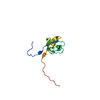
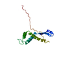
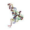
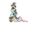
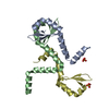
| Title | LIGAND BINDING DOMAIN OF HUMAN TRKB RECEPTOR | ||||||
 Components Components | PROTEIN (Brain Derived Neurotrophic Factor Receptor TrkB) | ||||||
 Keywords Keywords | TRANSFERASE / TRK RECEPTOR / RECEPTOR TYROSINE KINASE / 3D-DOMAIN SWAPPING | ||||||
| Function / homology |  Function and homology information Function and homology informationbrain-derived neurotrophic factor binding / brain-derived neurotrophic factor receptor activity / BDNF activates NTRK2 (TRKB) signaling / NTF4 activates NTRK2 (TRKB) signaling / NTF3 activates NTRK2 (TRKB) signaling / Activated NTRK2 signals through PLCG1 / peripheral nervous system neuron development / brain-derived neurotrophic factor receptor signaling pathway / retinal rod cell development / trans-synaptic signaling by BDNF, modulating synaptic transmission ...brain-derived neurotrophic factor binding / brain-derived neurotrophic factor receptor activity / BDNF activates NTRK2 (TRKB) signaling / NTF4 activates NTRK2 (TRKB) signaling / NTF3 activates NTRK2 (TRKB) signaling / Activated NTRK2 signals through PLCG1 / peripheral nervous system neuron development / brain-derived neurotrophic factor receptor signaling pathway / retinal rod cell development / trans-synaptic signaling by BDNF, modulating synaptic transmission / mechanoreceptor differentiation / neurotrophin binding / Activated NTRK2 signals through CDK5 / NTRK2 activates RAC1 / Activated NTRK2 signals through FYN / myelination in peripheral nervous system / Activated NTRK2 signals through PI3K / NGF-independant TRKA activation / regulation of GTPase activity / feeding behavior / glutamate secretion / neuronal action potential propagation / positive regulation of synapse assembly / positive regulation of axonogenesis / central nervous system neuron development / negative regulation of amyloid-beta formation / oligodendrocyte differentiation / negative regulation of anoikis / Activated NTRK2 signals through RAS / vasculogenesis / Activated NTRK2 signals through FRS2 and FRS3 / cellular response to brain-derived neurotrophic factor stimulus / cell surface receptor protein tyrosine kinase signaling pathway / axon terminus / learning / cellular response to amino acid stimulus / circadian rhythm / receptor protein-tyrosine kinase / positive regulation of neuron projection development / cerebral cortex development / neuron migration / Constitutive Signaling by Aberrant PI3K in Cancer / neuron differentiation / terminal bouton / long-term synaptic potentiation / protein autophosphorylation / PIP3 activates AKT signaling / PI5P, PP2A and IER3 Regulate PI3K/AKT Signaling / protease binding / early endosome membrane / dendritic spine / negative regulation of neuron apoptotic process / early endosome / positive regulation of MAPK cascade / positive regulation of phosphatidylinositol 3-kinase/protein kinase B signal transduction / signaling receptor complex / postsynaptic density / axon / positive regulation of cell population proliferation / positive regulation of gene expression / dendrite / perinuclear region of cytoplasm / protein homodimerization activity / ATP binding / plasma membrane / cytosol Similarity search - Function | ||||||
| Biological species |  Homo sapiens (human) Homo sapiens (human) | ||||||
| Method |  X-RAY DIFFRACTION / SYNCHROTRON / MOLECULAR REPLACEMENT / Resolution: 2.1 Å X-RAY DIFFRACTION / SYNCHROTRON / MOLECULAR REPLACEMENT / Resolution: 2.1 Å | ||||||
 Authors Authors | Wiesmann, C. / Ultsch, M.H. / Bass, S.H. / De Vos, A.M. | ||||||
 Citation Citation | Journal: J.Mol.Biol. / Year: 1999 Title: Crystal structures of the neurotrophin-binding domain of TrkA, TrkB and TrkC. Authors: Ultsch, M.H. / Wiesmann, C. / Simmons, L.C. / Henrich, J. / Yang, M. / Reilly, D. / Bass, S.H. / de Vos, A.M. | ||||||
| History |
|
- Structure visualization
| Structure viewer | Molecule: MolmilJmol/JSmol |
|---|
- Downloads & links
-Download
| PDBx/mmCIF format | 1wwb.cif.gz | 34.2 KB | Display | PDBx/mmCIF format |
|---|---|---|---|---|
| PDB format | pdb1wwb.ent.gz | 23.3 KB | Display | PDB format |
| PDBx/mmJSON format | 1wwb.json.gz | Tree view | PDBx/mmJSON format | |
| Others |  Other downloads Other downloads |
-Validation report
| Arichive directory | https://data.pdbj.org/pub/pdb/validation_reports/ww/1wwbftp://data.pdbj.org/pub/pdb/validation_reports/ww/1wwb | HTTPS FTP |
|---|
-Related structure data
| Related structure data |  1wwaC 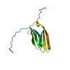 1wwcSC C: citing same article ( S: Starting model for refinement |
|---|---|
| Similar structure data |
-Links
 PDBj
PDBj


- Assembly
| Deposited unit | 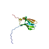
| ||||||||
|---|---|---|---|---|---|---|---|---|---|
| 1 |
| ||||||||
| Unit cell |
|
-Components
| #1: Protein | Mass: 11802.254 Da / Num. of mol.: 1 / Fragment: LIGAND BINDING DOMAIN Source method: isolated from a genetically manipulated source Source: (gene. exp.) Homo sapiens (human) / Production host:  |
|---|---|
| #2: Water | ChemComp-HOH /  Mass: 18.015 Da / Num. of mol.: 74 / Source method: isolated from a natural source / Formula: H2O Mass: 18.015 Da / Num. of mol.: 74 / Source method: isolated from a natural source / Formula: H2O |
| Has protein modification | Y |
-Experimental details
-Experiment
| Experiment | Method: X-RAY DIFFRACTION / Number of used crystals: 1 |
|---|
- Sample preparation
| Crystal | Density Matthews: 3.55 Å3/Da / Density % sol: 65.34 % | ||||||||||||||||||||||||||||||||||||||||||||||||||||||
|---|---|---|---|---|---|---|---|---|---|---|---|---|---|---|---|---|---|---|---|---|---|---|---|---|---|---|---|---|---|---|---|---|---|---|---|---|---|---|---|---|---|---|---|---|---|---|---|---|---|---|---|---|---|---|---|
| Crystal grow | pH: 8.5 / Details: pH 8.5 | ||||||||||||||||||||||||||||||||||||||||||||||||||||||
| Crystal grow | *PLUS Temperature: 4 ℃ / Method: vapor diffusion, sitting drop | ||||||||||||||||||||||||||||||||||||||||||||||||||||||
| Components of the solutions | *PLUS
|
-Data collection
| Diffraction | Mean temperature: 100 K |
|---|---|
| Diffraction source | Source: SYNCHROTRON / Site: CHESS  / Beamline: F1 / Wavelength: 0.91 / Beamline: F1 / Wavelength: 0.91 |
| Detector | Date: Mar 1, 1996 |
| Radiation | Protocol: SINGLE WAVELENGTH / Monochromatic (M) / Laue (L): M / Scattering type: x-ray |
| Radiation wavelength | Wavelength: 0.91 Å / Relative weight: 1 |
| Reflection | Resolution: 2.1→30 Å / Num. obs: 115960 / % possible obs: 98.7 % / Redundancy: 10.9 % / Rmerge(I) obs: 0.059 / Net I/σ(I): 7.3 |
| Reflection shell | Resolution: 2.1→2.18 Å / Rmerge(I) obs: 0.019 / % possible all: 97.3 |
- Processing
| Software |
| ||||||||||||||||||||||||||||||||||||||||||||||||||||||||||||||||||||||||||||||||
|---|---|---|---|---|---|---|---|---|---|---|---|---|---|---|---|---|---|---|---|---|---|---|---|---|---|---|---|---|---|---|---|---|---|---|---|---|---|---|---|---|---|---|---|---|---|---|---|---|---|---|---|---|---|---|---|---|---|---|---|---|---|---|---|---|---|---|---|---|---|---|---|---|---|---|---|---|---|---|---|---|---|
| Refinement | Method to determine structure: MOLECULAR REPLACEMENT Starting model: 1WWC Resolution: 2.1→20 Å / Rfactor Rfree error: 0.009 / Data cutoff high absF: 10000000 / Data cutoff low absF: 0.001 / Isotropic thermal model: RESTRAINED / Cross valid method: THROUGHOUT / σ(F): 0.2 Details: THE MOLECULE UNDERGOES 3D-DOMAIN SWAPPING, SUCH THAT STRAND A OF ONE MOLECULE REPLACES THE SAME STRAND A OF A SYMMETRY RELATED MATE
| ||||||||||||||||||||||||||||||||||||||||||||||||||||||||||||||||||||||||||||||||
| Refinement step | Cycle: LAST / Resolution: 2.1→20 Å
| ||||||||||||||||||||||||||||||||||||||||||||||||||||||||||||||||||||||||||||||||
| Refine LS restraints |
| ||||||||||||||||||||||||||||||||||||||||||||||||||||||||||||||||||||||||||||||||
| Software | *PLUS Name: X-PLOR / Version: 3.851 / Classification: refinement | ||||||||||||||||||||||||||||||||||||||||||||||||||||||||||||||||||||||||||||||||
| Refinement | *PLUS Highest resolution: 2.1 Å / σ(F): 0.2 / % reflection Rfree: 9.5 % / Rfactor obs: 0.247 | ||||||||||||||||||||||||||||||||||||||||||||||||||||||||||||||||||||||||||||||||
| Solvent computation | *PLUS | ||||||||||||||||||||||||||||||||||||||||||||||||||||||||||||||||||||||||||||||||
| Displacement parameters | *PLUS | ||||||||||||||||||||||||||||||||||||||||||||||||||||||||||||||||||||||||||||||||
| Refine LS restraints | *PLUS Type: x_angle_deg / Dev ideal: 1.9 |
