 Movie
Movie Controller

[English] 日本語
 Yorodumi
- PDB-1wve: p-Cresol Methylhydroxylase: Alteration of the Structure of the Fl... -
+ Open data

- Basic information
| Entry | Database: PDB / ID: 1wve | ||||||
|---|---|---|---|---|---|---|---|
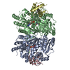
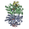
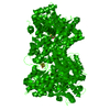
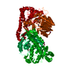
| Title | p-Cresol Methylhydroxylase: Alteration of the Structure of the Flavoprotein Subunit upon its Binding to the Cytochrome Subunit | ||||||
 Components Components | (4-cresol dehydrogenase [hydroxylating] ...) x 2 | ||||||
 Keywords Keywords | OXIDOREDUCTASE / FLAVOCYTOCHROME / ELECTRON-TRANSFER / FAD / HEME | ||||||
| Function / homology |  Function and homology information Function and homology information4-methylphenol dehydrogenase (hydroxylating) / 4-cresol dehydrogenase (hydroxylating) activity / lactate catabolic process / D-lactate dehydrogenase (cytochrome) activity / D-lactate dehydrogenase (NAD+) activity / FAD binding / electron transfer activity / heme binding / metal ion binding Similarity search - Function | ||||||
| Biological species |  Pseudomonas putida (bacteria) Pseudomonas putida (bacteria) | ||||||
| Method |  X-RAY DIFFRACTION / MOLECULAR REPLACEMENT / Resolution: 1.85 Å X-RAY DIFFRACTION / MOLECULAR REPLACEMENT / Resolution: 1.85 Å | ||||||
 Authors Authors | Cunane, L.M. / Chen, Z.-W. / McIntire, W.S. / Mathews, F.S. | ||||||
 Citation Citation | Journal: Biochemistry / Year: 2005 Title: p-Cresol Methylhydroxylase: Alteration of the Structure of the Flavoprotein Subunit upon Its Binding to the Cytochrome Subunit Authors: Cunane, L.M. / Chen, Z.-W. / McIntire, W.S. / Mathews, F.S. #1: Journal: J.Mol.Biol. / Year: 2000Title: Structures of the flavocytochrome p-cresol methylhydroxylase and its enzyme-substrate complex: gated substrate entry and proton relays support the proposed catalytic mechanism Authors: Cunane, L.M. / Chen, Z.-W. / Shamala, N. / Mathews, F.S. / Cronin, C.N. / McIntire, W.S. | ||||||
| History |
|
- Structure visualization
| Structure viewer | Molecule: MolmilJmol/JSmol |
|---|
- Downloads & links
-Download
| PDBx/mmCIF format | 1wve.cif.gz | 277.8 KB | Display | PDBx/mmCIF format |
|---|---|---|---|---|
| PDB format | pdb1wve.ent.gz | 217.3 KB | Display | PDB format |
| PDBx/mmJSON format | 1wve.json.gz | Tree view | PDBx/mmJSON format | |
| Others |  Other downloads Other downloads |
-Validation report
| Summary document | 1wve_validation.pdf.gz | 1.7 MB | Display | wwPDB validaton report |
|---|---|---|---|---|
| Full document | 1wve_full_validation.pdf.gz | 1.7 MB | Display | |
| Data in XML | 1wve_validation.xml.gz | 58.4 KB | Display | |
| Data in CIF | 1wve_validation.cif.gz | 84.5 KB | Display | |
| Arichive directory | https://data.pdbj.org/pub/pdb/validation_reports/wv/1wveftp://data.pdbj.org/pub/pdb/validation_reports/wv/1wve | HTTPS FTP |
-Related structure data
| Related structure data |  1wvfC  1diiS S: Starting model for refinement C: citing same article ( |
|---|---|
| Similar structure data |


-Links
 PDBj
PDBj


- Assembly
| Deposited unit | 
| ||||||||
|---|---|---|---|---|---|---|---|---|---|
| 1 |
| ||||||||
| Unit cell |
| ||||||||
| Details | The biological assembly is a heterotetramer. |
-Components
-4-cresol dehydrogenase [hydroxylating] ... , 2 types, 4 molecules ABCD
| #1: Protein | Mass: 57874.770 Da / Num. of mol.: 2 Source method: isolated from a genetically manipulated source Source: (gene. exp.) Pseudomonas putida (bacteria) / Production host: #2: Protein | Mass: 8611.642 Da / Num. of mol.: 2 Source method: isolated from a genetically manipulated source Source: (gene. exp.) Pseudomonas putida (bacteria) / Production host: |
|---|
-Non-polymers , 6 types, 1117 molecules 


| #3: Chemical |  Mass: 35.453 Da / Num. of mol.: 2 / Source method: obtained synthetically / Formula: Cl Mass: 35.453 Da / Num. of mol.: 2 / Source method: obtained synthetically / Formula: Cl#4: Chemical |  Mass: 785.550 Da / Num. of mol.: 2 / Source method: obtained synthetically / Formula: C27H33N9O15P2 / Comment: FAD*YM Mass: 785.550 Da / Num. of mol.: 2 / Source method: obtained synthetically / Formula: C27H33N9O15P2 / Comment: FAD*YM#5: Chemical | ChemComp-TRS /  Mass: 122.143 Da / Num. of mol.: 4 / Source method: obtained synthetically / Formula: C4H12NO3 / Comment: pH buffer*YM Mass: 122.143 Da / Num. of mol.: 4 / Source method: obtained synthetically / Formula: C4H12NO3 / Comment: pH buffer*YM#6: Chemical | ChemComp-ACY /  Mass: 60.052 Da / Num. of mol.: 4 / Source method: obtained synthetically / Formula: C2H4O2 Mass: 60.052 Da / Num. of mol.: 4 / Source method: obtained synthetically / Formula: C2H4O2#7: Chemical |  Mass: 616.487 Da / Num. of mol.: 2 / Source method: obtained synthetically / Formula: C34H32FeN4O4 Mass: 616.487 Da / Num. of mol.: 2 / Source method: obtained synthetically / Formula: C34H32FeN4O4#8: Water | ChemComp-HOH / | Mass: 18.015 Da / Num. of mol.: 1103 / Source method: isolated from a natural source / Formula: H2O |
|---|
-Details
| Has protein modification | Y |
|---|
-Experimental details
-Experiment
| Experiment | Method: X-RAY DIFFRACTION / Number of used crystals: 1 |
|---|
- Sample preparation
| Crystal | Density Matthews: 2.2 Å3/Da / Density % sol: 44 % |
|---|---|
| Crystal grow | Temperature: 296 K / Method: vapor diffusion, sitting drop / pH: 8.5 Details: PEG 8000, Tris, ammonium acetate, pH 8.5, VAPOR DIFFUSION, SITTING DROP, temperature 296K |
-Data collection
| Diffraction | Mean temperature: 100 K |
|---|---|
| Diffraction source | Source: ROTATING ANODE / Type: RIGAKU RU200 / Wavelength: 1.54 Å |
| Detector | Type: RIGAKU RAXIS IV / Detector: IMAGE PLATE / Date: Jun 3, 1998 |
| Radiation | Monochromator: Ni FILTER / Protocol: SINGLE WAVELENGTH / Monochromatic (M) / Laue (L): M / Scattering type: x-ray |
| Radiation wavelength | Wavelength: 1.54 Å / Relative weight: 1 |
| Reflection | Resolution: 1.85→30 Å / Num. all: 103498 / Num. obs: 94080 / % possible obs: 90.9 % / Observed criterion σ(F): 0 / Observed criterion σ(I): 0 / Redundancy: 5.1 % / Biso Wilson estimate: 18 Å2 / Rmerge(I) obs: 0.067 / Net I/σ(I): 18.6 |
| Reflection shell | Resolution: 1.85→1.92 Å / Redundancy: 1.5 % / Rmerge(I) obs: 0.285 / Mean I/σ(I) obs: 2.1 / % possible all: 52.3 |
- Processing
| Software |
| |||||||||||||||||||||||||||||||||||||||||||||||||
|---|---|---|---|---|---|---|---|---|---|---|---|---|---|---|---|---|---|---|---|---|---|---|---|---|---|---|---|---|---|---|---|---|---|---|---|---|---|---|---|---|---|---|---|---|---|---|---|---|---|---|
| Refinement | Method to determine structure: MOLECULAR REPLACEMENT Starting model: PDB code 1DII Resolution: 1.85→30 Å / Isotropic thermal model: Isotropic / Cross valid method: THROUGHOUT / σ(F): 0 / Stereochemistry target values: Engh & Huber
| |||||||||||||||||||||||||||||||||||||||||||||||||
| Displacement parameters | Biso mean: 22.8 Å2 | |||||||||||||||||||||||||||||||||||||||||||||||||
| Refine analyze |
| |||||||||||||||||||||||||||||||||||||||||||||||||
| Refinement step | Cycle: LAST / Resolution: 1.85→30 Å
| |||||||||||||||||||||||||||||||||||||||||||||||||
| Refine LS restraints |
| |||||||||||||||||||||||||||||||||||||||||||||||||
| LS refinement shell |
|
