 Movie
Movie Controller

[English] 日本語
 Yorodumi
- PDB-1diq: CRYSTAL STRUCTURE OF P-CRESOL METHYLHYDROXYLASE WITH SUBSTRATE BOUND -
+ Open data

- Basic information
| Entry | Database: PDB / ID: 1diq | |||||||||
|---|---|---|---|---|---|---|---|---|---|---|
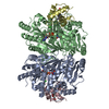
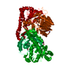
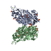
| Title | CRYSTAL STRUCTURE OF P-CRESOL METHYLHYDROXYLASE WITH SUBSTRATE BOUND | |||||||||
 Components Components | (P-CRESOL METHYLHYDROXYLASE) x 2 | |||||||||
 Keywords Keywords | OXIDOREDUCTASE / FLAVOCYTOCHROME / ELECTRON-TRANSFER / FAD / HEME / P-CRESOL | |||||||||
| Function / homology |  Function and homology information Function and homology information4-methylphenol dehydrogenase (hydroxylating) / 4-cresol dehydrogenase (hydroxylating) activity / lactate catabolic process / D-lactate dehydrogenase (cytochrome) activity / D-lactate dehydrogenase (NAD+) activity / FAD binding / electron transfer activity / heme binding / metal ion binding Similarity search - Function | |||||||||
| Biological species |  Pseudomonas putida (bacteria) Pseudomonas putida (bacteria) | |||||||||
| Method |  X-RAY DIFFRACTION / Resolution: 2.75 Å X-RAY DIFFRACTION / Resolution: 2.75 Å | |||||||||
 Authors Authors | Cunane, L.M. / Chen, Z.W. / Shamala, N. / Mathews, F.S. / Cronin, C.S. / McIntire, W.S. | |||||||||
 Citation Citation | Journal: J.Mol.Biol. / Year: 2000 Title: Structures of the flavocytochrome p-cresol methylhydroxylase and its enzyme-substrate complex: gated substrate entry and proton relays support the proposed catalytic mechanism. Authors: Cunane, L.M. / Chen, Z.W. / Shamala, N. / Mathews, F.S. / Cronin, C.N. / McIntire, W.S. #1: Journal: Biochemistry / Year: 1991Title: Three-dimensional Structure of p-Cresol Methylhydroxylase (Flavocytochrome c) from Pseudomonas putida at 3.0 A Resolution Authors: Mathews, F.S. / Chen, Z.W. / Bellamy, H. / McIntire, W.S. | |||||||||
| History |
|
- Structure visualization
| Structure viewer | Molecule: MolmilJmol/JSmol |
|---|
- Downloads & links
-Download
| PDBx/mmCIF format | 1diq.cif.gz | 252.4 KB | Display | PDBx/mmCIF format |
|---|---|---|---|---|
| PDB format | pdb1diq.ent.gz | 202.2 KB | Display | PDB format |
| PDBx/mmJSON format | 1diq.json.gz | Tree view | PDBx/mmJSON format | |
| Others |  Other downloads Other downloads |
-Validation report
| Arichive directory | https://data.pdbj.org/pub/pdb/validation_reports/di/1diqftp://data.pdbj.org/pub/pdb/validation_reports/di/1diq | HTTPS FTP |
|---|
-Related structure data


-Links
 PDBj
PDBj


- Assembly
| Deposited unit | 
| ||||||||
|---|---|---|---|---|---|---|---|---|---|
| 1 |
| ||||||||
| Unit cell |
| ||||||||
| Details | Asymmetric unit contains flavoprotein dimer related by molecular 2-fold axis. Two cytochrome subunits located on periphery of flavoprotein dimer. |
-Components
-Protein , 2 types, 4 molecules ABCD
| #1: Protein | Mass: 58005.965 Da / Num. of mol.: 2 / Fragment: FLAVOPROTEIN SUBUNIT / Source method: isolated from a natural source / Source: (natural) Pseudomonas putida (bacteria) / Cellular location: PERIPLASM / Strain: NCIMB 9869 / References: UniProt: P09788, EC: 1.17.99.1#2: Protein | Mass: 8611.642 Da / Num. of mol.: 2 / Fragment: CYTOCHROME SUBUNIT / Source method: isolated from a natural source / Source: (natural) Pseudomonas putida (bacteria) / Cellular location: PERIPLASM / Strain: NCIMB 9869 / References: UniProt: P09787, EC: 1.17.99.1 |
|---|
-Non-polymers , 5 types, 350 molecules 


| #3: Chemical |  Mass: 35.453 Da / Num. of mol.: 2 / Source method: obtained synthetically / Formula: Cl Mass: 35.453 Da / Num. of mol.: 2 / Source method: obtained synthetically / Formula: Cl#4: Chemical |  Mass: 785.550 Da / Num. of mol.: 2 / Source method: obtained synthetically / Formula: C27H33N9O15P2 / Comment: FAD*YM Mass: 785.550 Da / Num. of mol.: 2 / Source method: obtained synthetically / Formula: C27H33N9O15P2 / Comment: FAD*YM#5: Chemical |  Mass: 108.138 Da / Num. of mol.: 2 / Source method: obtained synthetically / Formula: C7H8O Mass: 108.138 Da / Num. of mol.: 2 / Source method: obtained synthetically / Formula: C7H8O#6: Chemical |  Mass: 618.503 Da / Num. of mol.: 2 / Source method: obtained synthetically / Formula: C34H34FeN4O4 Mass: 618.503 Da / Num. of mol.: 2 / Source method: obtained synthetically / Formula: C34H34FeN4O4#7: Water | ChemComp-HOH / | Mass: 18.015 Da / Num. of mol.: 342 / Source method: isolated from a natural source / Formula: H2O |
|---|
-Details
| Has protein modification | Y |
|---|
-Experimental details
-Experiment
| Experiment | Method: X-RAY DIFFRACTION / Number of used crystals: 1 |
|---|
- Sample preparation
| Crystal | Density Matthews: 2.53 Å3/Da / Density % sol: 51.43 % | ||||||||||||||||||||||||||||||
|---|---|---|---|---|---|---|---|---|---|---|---|---|---|---|---|---|---|---|---|---|---|---|---|---|---|---|---|---|---|---|---|
| Crystal grow | Temperature: 298 K / Method: liquid diffusion / pH: 7 Details: PEG 8000, Na/K phosphate, NaCl, Crystals soaked with substrate p-cresol , pH 7.0, LIQUID DIFFUSION, temperature 298K | ||||||||||||||||||||||||||||||
| Crystal | *PLUS Density % sol: 58 % | ||||||||||||||||||||||||||||||
| Crystal grow | *PLUS Method: vapor diffusion, hanging drop / Details: or interface diffusion | ||||||||||||||||||||||||||||||
| Components of the solutions | *PLUS
|
-Data collection
| Diffraction | Mean temperature: 298 K |
|---|---|
| Diffraction source | Source: ROTATING ANODE / Type: RIGAKU RU200 / Wavelength: 1.5418 |
| Detector | Type: SDMS / Detector: AREA DETECTOR / Date: Mar 17, 1990 |
| Radiation | Protocol: SINGLE WAVELENGTH / Monochromatic (M) / Laue (L): M / Scattering type: x-ray |
| Radiation wavelength | Wavelength: 1.5418 Å / Relative weight: 1 |
| Reflection | Resolution: 2.75→25 Å / Num. all: 35945 / Num. obs: 28932 / % possible obs: 80.5 % / Observed criterion σ(I): 2 / Redundancy: 5.5 % / Biso Wilson estimate: 47.4 Å2 / Rmerge(I) obs: 0.041 / Net I/σ(I): 16.5 |
| Reflection shell | Resolution: 2.75→2.95 Å / Redundancy: 2.4 % / Rmerge(I) obs: 0.106 / % possible all: 65.5 |
| Reflection shell | *PLUS % possible obs: 65.5 % |
- Processing
| Software |
| |||||||||||||||||||||||||
|---|---|---|---|---|---|---|---|---|---|---|---|---|---|---|---|---|---|---|---|---|---|---|---|---|---|---|
| Refinement | Resolution: 2.75→25 Å / σ(F): 0 / Stereochemistry target values: Engh & Huber
| |||||||||||||||||||||||||
| Refinement step | Cycle: LAST / Resolution: 2.75→25 Å
| |||||||||||||||||||||||||
| Refine LS restraints |
| |||||||||||||||||||||||||
| Software | *PLUS Name: 'CNS' / Classification: refinement | |||||||||||||||||||||||||
| Refine LS restraints | *PLUS
| |||||||||||||||||||||||||
| LS refinement shell | *PLUS Highest resolution: 2.75 Å / Lowest resolution: 3 Å / Rfactor Rfree: 0.289 / Rfactor Rwork: 0.21 |
