 Movie
Movie Controller

[English] 日本語
 Yorodumi
- PDB-1wv8: Crystal structure of hypothetical protein TTHA1013 from an extrem... -
+ Open data

- Basic information
| Entry | Database: PDB / ID: 1wv8 | ||||||
|---|---|---|---|---|---|---|---|
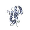
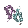
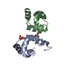
| Title | Crystal structure of hypothetical protein TTHA1013 from an extremely thermophilic bacterium thermus thermophilus HB8 | ||||||
 Components Components | hypothetical protein TTHA1013 | ||||||
 Keywords Keywords | STRUCTURAL GENOMICS / UNKNOWN FUNCTION / Hypothetical / novel fold / RIKEN Structural Genomics/Proteomics Initiative / RSGI | ||||||
| Function / homology | TTHA1013-like / Domain of unknown function DUF1902 / Domain of unknown function (DUF1902) / TTHA1013/TTHA0281-like / TTHA1013/TTHA0281-like / 2-Layer Sandwich / Alpha Beta / DUF1902 domain-containing protein Function and homology information Function and homology information | ||||||
| Biological species |   Thermus thermophilus (bacteria) Thermus thermophilus (bacteria) | ||||||
| Method |  X-RAY DIFFRACTION / SYNCHROTRON / MAD / Resolution: 2.2 Å X-RAY DIFFRACTION / SYNCHROTRON / MAD / Resolution: 2.2 Å | ||||||
 Authors Authors | Mizohata, E. / Hattori, M. / Kuramitsu, S. / Shirouzu, M. / Yokoyama, S. / RIKEN Structural Genomics/Proteomics Initiative (RSGI) | ||||||
 Citation Citation | Journal: Proteins / Year: 2005 Title: Crystal structure of the hypothetical protein TTHA1013 from Thermus thermophilus HB8 Authors: Hattori, M. / Mizohata, E. / Manzoku, M. / Bessho, Y. / Murayama, K. / Terada, T. / Kuramitsu, S. / Shirouzu, M. / Yokoyama, S. | ||||||
| History |
|
- Structure visualization
| Structure viewer | Molecule: MolmilJmol/JSmol |
|---|
- Downloads & links
-Download
| PDBx/mmCIF format | 1wv8.cif.gz | 26.7 KB | Display | PDBx/mmCIF format |
|---|---|---|---|---|
| PDB format | pdb1wv8.ent.gz | 17.2 KB | Display | PDB format |
| PDBx/mmJSON format | 1wv8.json.gz | Tree view | PDBx/mmJSON format | |
| Others |  Other downloads Other downloads |
-Validation report
| Summary document | 1wv8_validation.pdf.gz | 414 KB | Display | wwPDB validaton report |
|---|---|---|---|---|
| Full document | 1wv8_full_validation.pdf.gz | 416.8 KB | Display | |
| Data in XML | 1wv8_validation.xml.gz | 6.2 KB | Display | |
| Data in CIF | 1wv8_validation.cif.gz | 7.2 KB | Display | |
| Arichive directory | https://data.pdbj.org/pub/pdb/validation_reports/wv/1wv8ftp://data.pdbj.org/pub/pdb/validation_reports/wv/1wv8 | HTTPS FTP |
-Related structure data
| Similar structure data | |
|---|---|
| Other databases |


-Links
 PDBj
PDBj- Assembly
| Deposited unit | 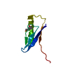
| |||||||||
|---|---|---|---|---|---|---|---|---|---|---|
| 1 | 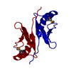
| |||||||||
| Unit cell |
| |||||||||
| Components on special symmetry positions |
|
-Components
| #1: Protein | Mass: 8013.877 Da / Num. of mol.: 1 Source method: isolated from a genetically manipulated source Source: (gene. exp.) Thermus thermophilus (bacteria) / Strain: HB8 / Plasmid: pET11a / Production host: |
|---|---|
| #2: Water | ChemComp-HOH /  Mass: 18.015 Da / Num. of mol.: 38 / Source method: isolated from a natural source / Formula: H2O Mass: 18.015 Da / Num. of mol.: 38 / Source method: isolated from a natural source / Formula: H2O |
| Has protein modification | Y |
-Experimental details
-Experiment
| Experiment | Method: X-RAY DIFFRACTION / Number of used crystals: 1 |
|---|
- Sample preparation
| Crystal | Density Matthews: 2.878 Å3/Da / Density % sol: 56 % |
|---|---|
| Crystal grow | Temperature: 293 K / Method: vapor diffusion, hanging drop / pH: 4.6 Details: ammonium sulfate, sodium acetate, PEG4000, Tris-HCl, Sodium chloride, pH 4.6, VAPOR DIFFUSION, HANGING DROP, temperature 293K |
-Data collection
| Diffraction | Mean temperature: 100 K | |||||||||||||||
|---|---|---|---|---|---|---|---|---|---|---|---|---|---|---|---|---|
| Diffraction source | Source: SYNCHROTRON / Site: SPring-8  / Beamline: BL26B1 / Wavelength: 0.96300, 0.97911, 0.97937, 1.00000 / Beamline: BL26B1 / Wavelength: 0.96300, 0.97911, 0.97937, 1.00000 | |||||||||||||||
| Detector | Type: RIGAKU / Detector: IMAGE PLATE / Date: Jun 17, 2004 | |||||||||||||||
| Radiation | Protocol: MAD / Monochromatic (M) / Laue (L): M / Scattering type: x-ray | |||||||||||||||
| Radiation wavelength |
| |||||||||||||||
| Reflection | Resolution: 2.2→50 Å / Num. obs: 5283 / Biso Wilson estimate: 30.3 Å2 |
- Processing
| Software |
| ||||||||||||||||||||||||||||||||||||||||||||||||||||||||||||
|---|---|---|---|---|---|---|---|---|---|---|---|---|---|---|---|---|---|---|---|---|---|---|---|---|---|---|---|---|---|---|---|---|---|---|---|---|---|---|---|---|---|---|---|---|---|---|---|---|---|---|---|---|---|---|---|---|---|---|---|---|---|
| Refinement | Method to determine structure: MAD / Resolution: 2.2→44.95 Å / Rfactor Rfree error: 0.016 / Data cutoff high absF: 573827.92 / Data cutoff low absF: 0 / Isotropic thermal model: RESTRAINED / Cross valid method: THROUGHOUT / σ(F): 0
| ||||||||||||||||||||||||||||||||||||||||||||||||||||||||||||
| Solvent computation | Solvent model: FLAT MODEL / Bsol: 51.5787 Å2 / ksol: 0.355245 e/Å3 | ||||||||||||||||||||||||||||||||||||||||||||||||||||||||||||
| Displacement parameters | Biso mean: 53.6 Å2
| ||||||||||||||||||||||||||||||||||||||||||||||||||||||||||||
| Refine analyze |
| ||||||||||||||||||||||||||||||||||||||||||||||||||||||||||||
| Refinement step | Cycle: LAST / Resolution: 2.2→44.95 Å
| ||||||||||||||||||||||||||||||||||||||||||||||||||||||||||||
| Refine LS restraints |
| ||||||||||||||||||||||||||||||||||||||||||||||||||||||||||||
| LS refinement shell | Resolution: 2.2→2.34 Å / Rfactor Rfree error: 0.055 / Total num. of bins used: 6
|
