 Movie
Movie Controller

[English] 日本語
 Yorodumi
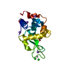
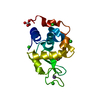
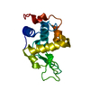
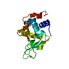
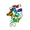
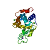
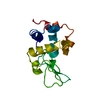
- PDB-1wtn: The structure of HEW Lysozyme Orthorhombic Crystal Growth under a... -
+ Open data

- Basic information
| Entry | Database: PDB / ID: 1wtn | ||||||
|---|---|---|---|---|---|---|---|
| Title | The structure of HEW Lysozyme Orthorhombic Crystal Growth under a High Magnetic Field | ||||||
 Components Components | Lysozyme C | ||||||
 Keywords Keywords | HYDROLASE / Allergen | ||||||
| Function / homology |  Function and homology information Function and homology informationLactose synthesis / Antimicrobial peptides / Neutrophil degranulation / beta-N-acetylglucosaminidase activity / cell wall macromolecule catabolic process / lysozyme / lysozyme activity / defense response to Gram-negative bacterium / killing of cells of another organism / defense response to Gram-positive bacterium ...Lactose synthesis / Antimicrobial peptides / Neutrophil degranulation / beta-N-acetylglucosaminidase activity / cell wall macromolecule catabolic process / lysozyme / lysozyme activity / defense response to Gram-negative bacterium / killing of cells of another organism / defense response to Gram-positive bacterium / defense response to bacterium / endoplasmic reticulum / extracellular space / identical protein binding / cytoplasm Similarity search - Function | ||||||
| Biological species |  | ||||||
| Method |  X-RAY DIFFRACTION / SYNCHROTRON / MOLECULAR REPLACEMENT / Resolution: 1.13 Å X-RAY DIFFRACTION / SYNCHROTRON / MOLECULAR REPLACEMENT / Resolution: 1.13 Å | ||||||
 Authors Authors | Saijo, S. / Yamada, Y. / Sato, T. / Tanaka, N. / Matsui, T. / Sazaki, G. / Nakajima, K. / Matsuura, Y. | ||||||
 Citation Citation | Journal: Acta Crystallogr.,Sect.D / Year: 2005 Title: Structural consequences of hen egg-white lysozyme orthorhombic crystal growth in a high magnetic field: validation of X-ray diffraction intensity, conformational energy searching and ...Title: Structural consequences of hen egg-white lysozyme orthorhombic crystal growth in a high magnetic field: validation of X-ray diffraction intensity, conformational energy searching and quantitative analysis of B factors and mosaicity. Authors: Saijo, S. / Yamada, Y. / Sato, T. / Tanaka, N. / Matsui, T. / Sazaki, G. / Nakajima, K. / Matsuura, Y. | ||||||
| History |
|
- Structure visualization
| Structure viewer | Molecule: MolmilJmol/JSmol |
|---|
- Downloads & links
-Download
| PDBx/mmCIF format | 1wtn.cif.gz | 40.1 KB | Display | PDBx/mmCIF format |
|---|---|---|---|---|
| PDB format | pdb1wtn.ent.gz | 27.8 KB | Display | PDB format |
| PDBx/mmJSON format | 1wtn.json.gz | Tree view | PDBx/mmJSON format | |
| Others |  Other downloads Other downloads |
-Validation report
| Arichive directory | https://data.pdbj.org/pub/pdb/validation_reports/wt/1wtnftp://data.pdbj.org/pub/pdb/validation_reports/wt/1wtn | HTTPS FTP |
|---|
-Related structure data


-Links
 PDBj
PDBj


- Assembly
| Deposited unit | 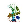
| ||||||||
|---|---|---|---|---|---|---|---|---|---|
| 1 |
| ||||||||
| Unit cell |
|
-Components
| #1: Protein | Mass: 14331.160 Da / Num. of mol.: 1 / Source method: isolated from a natural source / Source: (natural) |
|---|---|
| #2: Chemical | ChemComp-CL /   Mass: 35.453 Da / Num. of mol.: 1 / Source method: obtained synthetically / Formula: Cl Mass: 35.453 Da / Num. of mol.: 1 / Source method: obtained synthetically / Formula: Cl |
| #3: Water | ChemComp-HOH /  Mass: 18.015 Da / Num. of mol.: 116 / Source method: isolated from a natural source / Formula: H2O Mass: 18.015 Da / Num. of mol.: 116 / Source method: isolated from a natural source / Formula: H2O |
| Has protein modification | Y |
-Experimental details
-Experiment
| Experiment | Method: X-RAY DIFFRACTION / Number of used crystals: 8 |
|---|
- Sample preparation
| Crystal | Density Matthews: 2.27 Å3/Da / Density % sol: 45.81 % |
|---|---|
| Crystal grow | Temperature: 313 K / Method: under a high magnetic field of 10t / pH: 4.5 Details: NaCl, sodium acetate, pH 4.5, under a High Magnetic Field of 10T, temperature 313K |
-Data collection
| Diffraction |
| |||||||||||||||
|---|---|---|---|---|---|---|---|---|---|---|---|---|---|---|---|---|
| Diffraction source |
| |||||||||||||||
| Detector |
| |||||||||||||||
| Radiation | Monochromator: Si 111 / Protocol: SINGLE WAVELENGTH / Monochromatic (M) / Laue (L): M / Scattering type: x-ray | |||||||||||||||
| Radiation wavelength | Wavelength: 1 Å / Relative weight: 1 | |||||||||||||||
| Reflection | Resolution: 1.13→50 Å / Num. all: 34622 / Num. obs: 34449 / % possible obs: 99.5 % / Observed criterion σ(I): 3 | |||||||||||||||
| Reflection shell | Resolution: 1.13→1.16 Å / % possible all: 71.9 |

- Processing
| Software |
| ||||||||||||
|---|---|---|---|---|---|---|---|---|---|---|---|---|---|
| Refinement | Method to determine structure: MOLECULAR REPLACEMENT / Resolution: 1.13→16 Å / σ(F): 3 Details: technically defined here as 10 T and 0 T crystals, were grown by exposing them to a high magnetic field of 10 T (this entry 1WTN) and a control geomagnetic field of 40 uT(entry 1WTM), ...Details: technically defined here as 10 T and 0 T crystals, were grown by exposing them to a high magnetic field of 10 T (this entry 1WTN) and a control geomagnetic field of 40 uT(entry 1WTM), respectively (Sato,T., Yamada,Y., Saijo,S., Hori,T., Hirose,R., Tanaka,N., Sazaki,G., Nakajima,K., Igarashi,N., Tanaka,M. & Matsuura,Y. (2000). Acta Cryst. D56, 1079-1083.). A continuous homogeneous magnetic field of 10 T was produced by a liquid helium-free superconducting magnet (JMTD-10T100M9) (Japan Magnetic Technology Inc., Tokyo, Japan). Orthorhombic lysozyme crystals were grown, in the high magnetic field, via the batch crystallization method. The crystal growth conditions were 250 mg.ml-1 lysozyme, 42 mM NaCl in 50 mM sodium acetate buffer with pH 4.5 at 313 K; 11 days duration. The dimensions of the 10 T and 0 T crystals were typically 0.45 X 0.50 X 2.00 and 0.50 X 0.50 X 2.00 mm, respectively. The starting model for molecular replacement in each case was the refined atomic coordinates (entry 1BGI) of the HEW lysozyme obtained from the PDB (Oki,H., Matsuura,Y., Komatsu,H. & Chernov,A.A. (1999). Acta Cryst. D55, 114- 121.), i.e., our best resolution structure for the orthorhombic lysozyme refined so far (at 1.7 A resolution).
| ||||||||||||
| Refine analyze | Luzzati coordinate error obs: 0.16 Å | ||||||||||||
| Refinement step | Cycle: LAST / Resolution: 1.13→16 Å
| ||||||||||||
| Refine LS restraints |
|
