 ムービー
ムービー コントローラー


+ データを開く

- 基本情報
基本情報
| 登録情報 | データベース: PDB / ID: 1wtn | ||||||
|---|---|---|---|---|---|---|---|
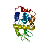
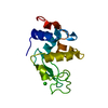
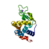
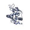
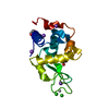
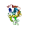
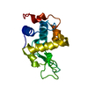
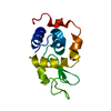
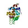
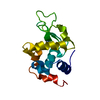
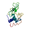
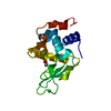
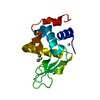
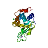
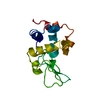
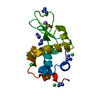
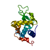
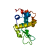
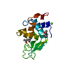
| タイトル | The structure of HEW Lysozyme Orthorhombic Crystal Growth under a High Magnetic Field | ||||||
 要素 要素 | Lysozyme C | ||||||
 キーワード キーワード |  HYDROLASE (加水分解酵素) / Allergen (アレルゲン) HYDROLASE (加水分解酵素) / Allergen (アレルゲン) | ||||||
| 機能・相同性 |  機能・相同性情報抗微生物ペプチド / Neutrophil degranulation / beta-N-acetylglucosaminidase activity / cell wall macromolecule catabolic process / リゾチーム / lysozyme activity / killing of cells of another organism / defense response to Gram-negative bacterium / defense response to Gram-positive bacterium / defense response to bacterium ...抗微生物ペプチド / Neutrophil degranulation / beta-N-acetylglucosaminidase activity / cell wall macromolecule catabolic process / リゾチーム / lysozyme activity / killing of cells of another organism / defense response to Gram-negative bacterium / defense response to Gram-positive bacterium / defense response to bacterium / 小胞体 / extracellular space / identical protein binding / 細胞質 機能・相同性情報抗微生物ペプチド / Neutrophil degranulation / beta-N-acetylglucosaminidase activity / cell wall macromolecule catabolic process / リゾチーム / lysozyme activity / killing of cells of another organism / defense response to Gram-negative bacterium / defense response to Gram-positive bacterium / defense response to bacterium ...抗微生物ペプチド / Neutrophil degranulation / beta-N-acetylglucosaminidase activity / cell wall macromolecule catabolic process / リゾチーム / lysozyme activity / killing of cells of another organism / defense response to Gram-negative bacterium / defense response to Gram-positive bacterium / defense response to bacterium / 小胞体 / extracellular space / identical protein binding / 細胞質類似検索 - 分子機能 | ||||||
| 生物種 |  Gallus gallus (ニワトリ) Gallus gallus (ニワトリ) | ||||||
| 手法 | X線回折 / シンクロトロン / 分子置換 / 解像度: 1.13 Å | ||||||
 データ登録者 データ登録者 | Saijo, S. / Yamada, Y. / Sato, T. / Tanaka, N. / Matsui, T. / Sazaki, G. / Nakajima, K. / Matsuura, Y. | ||||||
 引用 引用 | ジャーナル: Acta Crystallogr.,Sect.D / 年: 2005 タイトル: Structural consequences of hen egg-white lysozyme orthorhombic crystal growth in a high magnetic field: validation of X-ray diffraction intensity, conformational energy searching and ...タイトル: Structural consequences of hen egg-white lysozyme orthorhombic crystal growth in a high magnetic field: validation of X-ray diffraction intensity, conformational energy searching and quantitative analysis of B factors and mosaicity. 著者: Saijo, S. / Yamada, Y. / Sato, T. / Tanaka, N. / Matsui, T. / Sazaki, G. / Nakajima, K. / Matsuura, Y. | ||||||
| 履歴 |
|
- 構造の表示
構造の表示
| 構造ビューア | 分子: MolmilJmol/JSmol |
|---|
- ダウンロードとリンク
-ダウンロード
| PDBx/mmCIF形式 | 1wtn.cif.gz | 35.9 KB | 表示 | PDBx/mmCIF形式 |
|---|---|---|---|---|
| PDB形式 | pdb1wtn.ent.gz | 27.8 KB | 表示 | PDB形式 |
| PDBx/mmJSON形式 | 1wtn.json.gz | ツリー表示 | PDBx/mmJSON形式 | |
| その他 |  その他のダウンロード その他のダウンロード |
-検証レポート
| アーカイブディレクトリ | https://data.pdbj.org/pub/pdb/validation_reports/wt/1wtnftp://data.pdbj.org/pub/pdb/validation_reports/wt/1wtn | HTTPS FTP |
|---|
-関連構造データ


-リンク
 PDBj
PDBj


- 集合体
集合体
| 登録構造単位 | 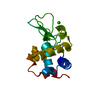
| ||||||||
|---|---|---|---|---|---|---|---|---|---|
| 1 |
| ||||||||
| 単位格子 |
|
-要素
| #1: タンパク質 | 分子量: 14331.160 Da / 分子数: 1 / 由来タイプ: 天然 / 由来: (天然) Gallus gallus (ニワトリ) / Cell: egg / 細胞内の位置: cytoplasm(white) / 参照: UniProt: P00698, リゾチーム |
|---|---|
| #2: 化合物 | ChemComp-CL / 塩化物  分子量: 35.453 Da / 分子数: 1 / 由来タイプ: 合成 / 式: Cl 分子量: 35.453 Da / 分子数: 1 / 由来タイプ: 合成 / 式: Cl |
| #3: 水 | ChemComp-HOH / 水 分子量: 18.015 Da / 分子数: 116 / 由来タイプ: 天然 / 式: H2O 分子量: 18.015 Da / 分子数: 116 / 由来タイプ: 天然 / 式: H2O |
-実験情報
-実験
| 実験 | 手法: X線回折 / 使用した結晶の数: 8 |
|---|
- 試料調製
試料調製
| 結晶 | マシュー密度: 2.27 Å3/Da / 溶媒含有率: 45.81 % |
|---|---|
| 結晶化 | 温度: 313 K / 手法: under a high magnetic field of 10t / pH: 4.5 詳細: NaCl, sodium acetate, pH 4.5, under a High Magnetic Field of 10T, temperature 313K |
-データ収集
| 回折 |
| |||||||||||||||
|---|---|---|---|---|---|---|---|---|---|---|---|---|---|---|---|---|
| 放射光源 |
| |||||||||||||||
| 検出器 |
| |||||||||||||||
| 放射 | モノクロメーター: Si 111 / プロトコル: SINGLE WAVELENGTH / 単色(M)・ラウエ(L): M / 散乱光タイプ: x-ray | |||||||||||||||
| 放射波長 | 波長: 1 Å / 相対比: 1 | |||||||||||||||
| 反射 | 解像度: 1.13→50 Å / Num. all: 34622 / Num. obs: 34449 / % possible obs: 99.5 % / Observed criterion σ(I): 3 | |||||||||||||||
| 反射 シェル | 解像度: 1.13→1.16 Å / % possible all: 71.9 |

- 解析
解析
| ソフトウェア |
| ||||||||||||
|---|---|---|---|---|---|---|---|---|---|---|---|---|---|
| 精密化 | 構造決定の手法: 分子置換 / 解像度: 1.13→16 Å / σ(F): 3 詳細: technically defined here as 10 T and 0 T crystals, were grown by exposing them to a high magnetic field of 10 T (this entry 1WTN) and a control geomagnetic field of 40 uT(entry 1WTM), ...詳細: technically defined here as 10 T and 0 T crystals, were grown by exposing them to a high magnetic field of 10 T (this entry 1WTN) and a control geomagnetic field of 40 uT(entry 1WTM), respectively (Sato,T., Yamada,Y., Saijo,S., Hori,T., Hirose,R., Tanaka,N., Sazaki,G., Nakajima,K., Igarashi,N., Tanaka,M. & Matsuura,Y. (2000). Acta Cryst. D56, 1079-1083.). A continuous homogeneous magnetic field of 10 T was produced by a liquid helium-free superconducting magnet (JMTD-10T100M9) (Japan Magnetic Technology Inc., Tokyo, Japan). Orthorhombic lysozyme crystals were grown, in the high magnetic field, via the batch crystallization method. The crystal growth conditions were 250 mg.ml-1 lysozyme, 42 mM NaCl in 50 mM sodium acetate buffer with pH 4.5 at 313 K; 11 days duration. The dimensions of the 10 T and 0 T crystals were typically 0.45 X 0.50 X 2.00 and 0.50 X 0.50 X 2.00 mm, respectively. The starting model for molecular replacement in each case was the refined atomic coordinates (entry 1BGI) of the HEW lysozyme obtained from the PDB (Oki,H., Matsuura,Y., Komatsu,H. & Chernov,A.A. (1999). Acta Cryst. D55, 114- 121.), i.e., our best resolution structure for the orthorhombic lysozyme refined so far (at 1.7 A resolution).
| ||||||||||||
| Refine analyze | Luzzati coordinate error obs: 0.16 Å | ||||||||||||
| 精密化ステップ | サイクル: LAST / 解像度: 1.13→16 Å
| ||||||||||||
| 拘束条件 |
|
