[English] 日本語
Yorodumi
- PDB-1wql: Cumene dioxygenase (cumA1A2) from Pseudomonas fluorescens IP01 -

+
Open data


ID or keywords:

Loading...

-
Basic information

Entry
Database: PDB / ID: 1wql
TitleCumene dioxygenase (cumA1A2) from Pseudomonas fluorescens IP01
Components
  • ethylbenzene dioxygenase small subunit
  • iron-sulfur protein large subunit of cumene dioxygenase
KeywordsOXIDOREDUCTASE / biphenyl dioxygenase / cumene dioxygenase / naphthalene dioxygenase / polychlorinated biphenyl degradation / Rieske non-heme iron dioxygenase
Function / homology
Function and homology information


: / dioxygenase activity / 2 iron, 2 sulfur cluster binding / iron ion binding
Similarity search - Function
Ring-hydroxylating dioxygenase, alpha subunit NdoB-like, C-terminal / Ring hydroxylating beta subunit / Ring-hydroxylating dioxygenase beta subunit / Aromatic-ring-hydroxylating dioxygenase, alpha subunit / Aromatic-ring-hydroxylating dioxygenase, alpha subunit, C-terminal domain / Ring hydroxylating alpha subunit (catalytic domain) / Aromatic-ring-hydroxylating dioxygenase, 2Fe-2S-binding site / Bacterial ring hydroxylating dioxygenases alpha-subunit signature. / Naphthalene 1,2-dioxygenase Alpha Subunit; Chain A, domain 1 / Naphthalene 1,2-dioxygenase Alpha Subunit; Chain A, domain 1 ...Ring-hydroxylating dioxygenase, alpha subunit NdoB-like, C-terminal / Ring hydroxylating beta subunit / Ring-hydroxylating dioxygenase beta subunit / Aromatic-ring-hydroxylating dioxygenase, alpha subunit / Aromatic-ring-hydroxylating dioxygenase, alpha subunit, C-terminal domain / Ring hydroxylating alpha subunit (catalytic domain) / Aromatic-ring-hydroxylating dioxygenase, 2Fe-2S-binding site / Bacterial ring hydroxylating dioxygenases alpha-subunit signature. / Naphthalene 1,2-dioxygenase Alpha Subunit; Chain A, domain 1 / Naphthalene 1,2-dioxygenase Alpha Subunit; Chain A, domain 1 / Rieske Iron-sulfur Protein / Rieske [2Fe-2S] iron-sulphur domain / 3-layer Sandwich / Nuclear Transport Factor 2; Chain: A, - #50 / Rieske [2Fe-2S] iron-sulphur domain / Rieske [2Fe-2S] domain / Rieske [2Fe-2S] iron-sulfur domain profile. / Rieske [2Fe-2S] iron-sulphur domain superfamily / NTF2-like domain superfamily / Nuclear Transport Factor 2; Chain: A, / Roll / Alpha-Beta Complex / Mainly Beta / Alpha Beta
Similarity search - Domain/homology
: / FE2/S2 (INORGANIC) CLUSTER / OXYGEN MOLECULE / Iron-sulfur protein large subunit of cumene dioxygenase / Ethylbenzene dioxygenase small subunit
Similarity search - Component
Biological speciesPseudomonas fluorescens (bacteria)
MethodX-RAY DIFFRACTION / SYNCHROTRON / MOLECULAR REPLACEMENT / Resolution: 2.2 Å
AuthorsDong, X. / Fushinobu, S. / Fukuda, E. / Terada, T. / Nakamura, S. / Shimizu, K. / Nojiri, H. / Omori, T. / Shoun, H. / Wakagi, T.
CitationJournal: J.BACTERIOL. / Year: 2005
Title: Crystal Structure of the Terminal Oxygenase Component of Cumene Dioxygenase from Pseudomonas fluorescens IP01
Authors: Dong, X. / Fushinobu, S. / Fukuda, E. / Terada, T. / Nakamura, S. / Shimizu, K. / Nojiri, H. / Omori, T. / Shoun, H. / Wakagi, T.
History
DepositionSep 30, 2004Deposition site: PDBJ / Processing site: PDBJ
Revision 1.0Mar 29, 2005Provider: repository / Type: Initial release
Revision 1.1Apr 30, 2008Group: Version format compliance
Revision 1.2Jul 13, 2011Group: Derived calculations / Version format compliance
Revision 1.3Oct 25, 2023Group: Data collection / Database references ...Data collection / Database references / Derived calculations / Refinement description
Category: chem_comp_atom / chem_comp_bond ...chem_comp_atom / chem_comp_bond / database_2 / pdbx_initial_refinement_model / pdbx_struct_conn_angle / struct_conn / struct_site
Item: _database_2.pdbx_DOI / _database_2.pdbx_database_accession ..._database_2.pdbx_DOI / _database_2.pdbx_database_accession / _pdbx_struct_conn_angle.ptnr1_auth_comp_id / _pdbx_struct_conn_angle.ptnr1_auth_seq_id / _pdbx_struct_conn_angle.ptnr1_label_atom_id / _pdbx_struct_conn_angle.ptnr1_label_comp_id / _pdbx_struct_conn_angle.ptnr1_label_seq_id / _pdbx_struct_conn_angle.ptnr2_label_atom_id / _pdbx_struct_conn_angle.ptnr3_auth_comp_id / _pdbx_struct_conn_angle.ptnr3_auth_seq_id / _pdbx_struct_conn_angle.ptnr3_label_atom_id / _pdbx_struct_conn_angle.ptnr3_label_comp_id / _pdbx_struct_conn_angle.ptnr3_label_seq_id / _pdbx_struct_conn_angle.value / _struct_conn.pdbx_dist_value / _struct_conn.ptnr1_auth_comp_id / _struct_conn.ptnr1_auth_seq_id / _struct_conn.ptnr1_label_asym_id / _struct_conn.ptnr1_label_atom_id / _struct_conn.ptnr1_label_comp_id / _struct_conn.ptnr1_label_seq_id / _struct_conn.ptnr2_auth_comp_id / _struct_conn.ptnr2_auth_seq_id / _struct_conn.ptnr2_label_asym_id / _struct_conn.ptnr2_label_atom_id / _struct_conn.ptnr2_label_comp_id / _struct_conn.ptnr2_label_seq_id / _struct_site.pdbx_auth_asym_id / _struct_site.pdbx_auth_comp_id / _struct_site.pdbx_auth_seq_id

-
Structure visualization

Structure viewerMolecule:
MolmilJmol/JSmol

Downloads & links

-
Assembly

Deposited unit
A: iron-sulfur protein large subunit of cumene dioxygenase
B: ethylbenzene dioxygenase small subunit
hetero molecules


Theoretical massNumber of molelcules
Total (without water)74,2625
Polymers73,9982
Non-polymers2643
Water8,503472
1
A: iron-sulfur protein large subunit of cumene dioxygenase
B: ethylbenzene dioxygenase small subunit
hetero molecules

A: iron-sulfur protein large subunit of cumene dioxygenase
B: ethylbenzene dioxygenase small subunit
hetero molecules

A: iron-sulfur protein large subunit of cumene dioxygenase
B: ethylbenzene dioxygenase small subunit
hetero molecules


Theoretical massNumber of molelcules
Total (without water)222,78615
Polymers221,9956
Non-polymers7919
Water1086
TypeNameSymmetry operationNumber
identity operation1_555x,y,z1
crystal symmetry operation5_555z,x,y1
crystal symmetry operation9_555y,z,x1
Buried area32760 Å2
ΔGint-200 kcal/mol
Surface area56450 Å2
MethodPISA, PQS
Unit cell
Length a, b, c (Å)147.036, 147.036, 147.036
Angle α, β, γ (deg.)90.00, 90.00, 90.00
Int Tables number198
Space group name H-MP213
Components on special symmetry positions
IDModelComponents
11B-278-

HOH

Details
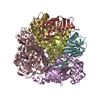
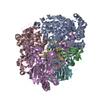
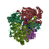
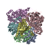
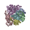
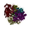
The biological assembly is a hexamer generated from the dimer in the asymmetric unit by the operations: z, x, y and y, z, x.

-
Components

-
Protein , 2 types, 2 molecules AB

#1: Protein iron-sulfur protein large subunit of cumene dioxygenase / / cumene dioxygenase large subunit


Mass: 52357.992 Da / Num. of mol.: 1
Source method: isolated from a genetically manipulated source
Source: (gene. exp.) Pseudomonas fluorescens (bacteria) / Gene: CUMA1 / Plasmid: pUC118 / Species (production host): Escherichia coli / Production host: Escherichia coli BL21(DE3) (bacteria) / Strain (production host): BL21 (DE3)
References: UniProt: Q51743, Oxidoreductases; Acting on paired donors, with incorporation or reduction of molecular oxygen; With NADH or NADPH as one donor, and incorporation of two atoms of oxygen into the other donor
#2: Protein ethylbenzene dioxygenase small subunit / cumene dioxygenase small subunit


Mass: 21640.428 Da / Num. of mol.: 1
Source method: isolated from a genetically manipulated source
Source: (gene. exp.) Pseudomonas fluorescens (bacteria) / Gene: CUMA2 / Plasmid: pUC118 / Species (production host): Escherichia coli / Production host: Escherichia coli BL21(DE3) (bacteria) / Strain (production host): BL21 (DE3)
References: UniProt: Q51744, Oxidoreductases; Acting on paired donors, with incorporation or reduction of molecular oxygen; With NADH or NADPH as one donor, and incorporation of two atoms of oxygen into the other donor

-
Non-polymers , 4 types, 475 molecules

#3: Chemical ChemComp-FE2 / FE (II) ION


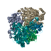
Mass: 55.845 Da / Num. of mol.: 1 / Source method: obtained synthetically / Formula: Fe
#4: Chemical ChemComp-FES / FE2/S2 (INORGANIC) CLUSTER / Iron–sulfur cluster


Mass: 175.820 Da / Num. of mol.: 1 / Source method: obtained synthetically / Formula: Fe2S2
#5: Chemical ChemComp-OXY / OXYGEN MOLECULE / Oxygen


Mass: 31.999 Da / Num. of mol.: 1 / Source method: obtained synthetically / Formula: O2
#6: Water ChemComp-HOH / water / Water


Mass: 18.015 Da / Num. of mol.: 472 / Source method: isolated from a natural source / Formula: H2O

-
Experimental details

-
Experiment

ExperimentMethod: X-RAY DIFFRACTION / Number of used crystals: 1

-
Sample preparation

CrystalDensity Matthews: 3.5 Å3/Da / Density % sol: 65 %
Crystal growTemperature: 298 K / Method: vapor diffusion, sitting drop / pH: 8.1
Details: PEG 3350, tri-lithium citrate tetrahydrate, pH 8.1, VAPOR DIFFUSION, SITTING DROP, temperature 298K

-
Data collection

DiffractionMean temperature: 100 K
Diffraction sourceSource: SYNCHROTRON / Site: SPring-8 / Beamline: BL40B2 / Wavelength: 0.9794 Å
DetectorType: ADSC QUANTUM 4 / Detector: CCD / Date: Oct 18, 2002
RadiationMonochromator: Si(111) / Protocol: SINGLE WAVELENGTH / Monochromatic (M) / Laue (L): M / Scattering type: x-ray
Radiation wavelengthWavelength: 0.9794 Å / Relative weight: 1
ReflectionResolution: 2.2→20 Å / Num. all: 53921 / Num. obs: 53807 / % possible obs: 99.8 % / Observed criterion σ(F): 0 / Observed criterion σ(I): 0 / Redundancy: 15.5 % / Biso Wilson estimate: 12.8 Å2 / Rsym value: 0.113 / Net I/σ(I): 17.7
Reflection shellResolution: 2.2→2.28 Å / Mean I/σ(I) obs: 6.5 / Num. unique all: 5317 / Rsym value: 0.295 / % possible all: 100

-
Processing

Software
NameVersionClassification
CNS1.1refinement
HKL-2000data reduction
SCALEPACKdata scaling
MOLREPphasing
RefinementMethod to determine structure: MOLECULAR REPLACEMENT
Starting model: PDB ENTRY 1EG9
Resolution: 2.2→19.65 Å / Rfactor Rfree error: 0.004 / Data cutoff high absF: 3753443.73 / Data cutoff low absF: 0 / Isotropic thermal model: RESTRAINED / Cross valid method: THROUGHOUT / σ(F): 0 / Stereochemistry target values: Engh & Huber
RfactorNum. reflection% reflectionSelection details
Rfree0.196 2678 5 %RANDOM
Rwork0.173 ---
all0.174 53807 --
obs0.173 53762 99.8 %-
Solvent computationSolvent model: FLAT MODEL / Bsol: 39.7029 Å2 / ksol: 0.373348 e/Å3
Displacement parametersBiso mean: 21.8 Å2
Baniso -1Baniso -2Baniso -3
1-0 Å20 Å20 Å2
2--0 Å20 Å2
3---0 Å2
Refine analyze
FreeObs
Luzzati coordinate error0.24 Å0.21 Å
Luzzati d res low-5 Å
Luzzati sigma a0.17 Å0.09 Å
Refinement stepCycle: LAST / Resolution: 2.2→19.65 Å
ProteinNucleic acidLigandSolventTotal
Num. atoms4994 0 7 472 5473
Refine LS restraints
Refine-IDTypeDev idealDev ideal target
X-RAY DIFFRACTIONc_bond_d0.006
X-RAY DIFFRACTIONc_angle_deg1.4
X-RAY DIFFRACTIONc_dihedral_angle_d23.6
X-RAY DIFFRACTIONc_improper_angle_d0.75
X-RAY DIFFRACTIONc_mcbond_it1.671.5
X-RAY DIFFRACTIONc_mcangle_it2.332
X-RAY DIFFRACTIONc_scbond_it2.792
X-RAY DIFFRACTIONc_scangle_it3.732.5
LS refinement shellResolution: 2.2→2.34 Å / Rfactor Rfree error: 0.01 / Total num. of bins used: 6
RfactorNum. reflection% reflection
Rfree0.221 450 5.1 %
Rwork0.183 8445 -
obs--100 %
Xplor file
Refine-IDSerial noParam fileTopol file
X-RAY DIFFRACTION1PROTEIN_REP.PARAMPROTEIN.TOP
X-RAY DIFFRACTION2WATER_REP.PARAMWATER.TOP
X-RAY DIFFRACTION3ION.PARAMION.TOP
X-RAY DIFFRACTION4FESO2.PARAMFESO2.TOP

+
About Yorodumi

-
News

-
Feb 9, 2022. New format data for meta-information of EMDB entries

New format data for meta-information of EMDB entries

  • Version 3 of the EMDB header file is now the official format.
  • The previous official version 1.9 will be removed from the archive.

Related info.:EMDB header

External links:wwPDB to switch to version 3 of the EMDB data model

-
Aug 12, 2020. Covid-19 info

Covid-19 info

URL: https://pdbj.org/emnavi/covid19.php

New page: Covid-19 featured information page in EM Navigator.

Related info.:Covid-19 info / Mar 5, 2020. Novel coronavirus structure data

+
Mar 5, 2020. Novel coronavirus structure data

Novel coronavirus structure data

Related info.:Yorodumi Speices / Aug 12, 2020. Covid-19 info

External links:COVID-19 featured content - PDBj / Molecule of the Month (242):Coronavirus Proteases

+
Jan 31, 2019. EMDB accession codes are about to change! (news from PDBe EMDB page)

EMDB accession codes are about to change! (news from PDBe EMDB page)

  • The allocation of 4 digits for EMDB accession codes will soon come to an end. Whilst these codes will remain in use, new EMDB accession codes will include an additional digit and will expand incrementally as the available range of codes is exhausted. The current 4-digit format prefixed with “EMD-” (i.e. EMD-XXXX) will advance to a 5-digit format (i.e. EMD-XXXXX), and so on. It is currently estimated that the 4-digit codes will be depleted around Spring 2019, at which point the 5-digit format will come into force.
  • The EM Navigator/Yorodumi systems omit the EMD- prefix.

Related info.:Q: What is EMD? / ID/Accession-code notation in Yorodumi/EM Navigator

External links:EMDB Accession Codes are Changing Soon! / Contact to PDBj

+
Jul 12, 2017. Major update of PDB

Major update of PDB

  • wwPDB released updated PDB data conforming to the new PDBx/mmCIF dictionary.
  • This is a major update changing the version number from 4 to 5, and with Remediation, in which all the entries are updated.
  • In this update, many items about electron microscopy experimental information are reorganized (e.g. em_software).
  • Now, EM Navigator and Yorodumi are based on the updated data.

External links:wwPDB Remediation / Enriched Model Files Conforming to OneDep Data Standards Now Available in the PDB FTP Archive

-
Yorodumi

Thousand views of thousand structures

  • Yorodumi is a browser for structure data from EMDB, PDB, SASBDB, etc.
  • This page is also the successor to EM Navigator detail page, and also detail information page/front-end page for Omokage search.
  • The word "yorodu" (or yorozu) is an old Japanese word meaning "ten thousand". "mi" (miru) is to see.

Related info.:EMDB / PDB / SASBDB / Comparison of 3 databanks / Yorodumi Search / Aug 31, 2016. New EM Navigator & Yorodumi / Yorodumi Papers / Jmol/JSmol / Function and homology information / Changes in new EM Navigator and Yorodumi

Read more