 Movie
Movie Controller

[English] 日本語
 Yorodumi
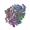
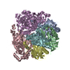
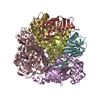
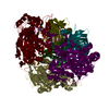
- PDB-2xsh: CRYSTAL STRUCTURE OF P4 VARIANT OF BIPHENYL DIOXYGENASE FROM BURK... -
+ Open data

- Basic information
| Entry | Database: PDB / ID: 2xsh | ||||||
|---|---|---|---|---|---|---|---|
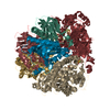
| Title | CRYSTAL STRUCTURE OF P4 VARIANT OF BIPHENYL DIOXYGENASE FROM BURKHOLDERIA XENOVORANS LB400 IN COMPLEX WITH 2,6 DI CHLOROBIPHENYL | ||||||
 Components Components | (BIPHENYL DIOXYGENASE SUBUNIT ...) x 2 | ||||||
 Keywords Keywords | OXIDOREDUCTASE | ||||||
| Function / homology |  Function and homology information Function and homology informationbiphenyl 2,3-dioxygenase / biphenyl 2,3-dioxygenase activity / 3-phenylpropionate catabolic process / 2 iron, 2 sulfur cluster binding / iron ion binding Similarity search - Function | ||||||
| Biological species |  BURKHOLDERIA XENOVORANS (bacteria) BURKHOLDERIA XENOVORANS (bacteria) | ||||||
| Method |  X-RAY DIFFRACTION / SYNCHROTRON / MOLECULAR REPLACEMENT / Resolution: 2.29 Å X-RAY DIFFRACTION / SYNCHROTRON / MOLECULAR REPLACEMENT / Resolution: 2.29 Å | ||||||
 Authors Authors | Kumar, P. / Bolin, J.T. | ||||||
 Citation Citation | Journal: J.Mol.Biol. / Year: 2011 Title: Structural Insight Into the Expanded Pcb-Degrading Abilities of a Biphenyl Dioxygenase Obtained by Directed Evolution. Authors: Kumar, P. / Mohammadi, M. / Viger, J.F. / Barriault, D. / Gomez-Gil, L. / Eltis, L.D. / Bolin, J.T. / Sylvestre, M. | ||||||
| History |
|
- Structure visualization
| Structure viewer | Molecule: MolmilJmol/JSmol |
|---|
- Downloads & links
-Download
| PDBx/mmCIF format | 2xsh.cif.gz | 1.4 MB | Display | PDBx/mmCIF format |
|---|---|---|---|---|
| PDB format | pdb2xsh.ent.gz | 1.2 MB | Display | PDB format |
| PDBx/mmJSON format | 2xsh.json.gz | Tree view | PDBx/mmJSON format | |
| Others |  Other downloads Other downloads |
-Validation report
| Arichive directory | https://data.pdbj.org/pub/pdb/validation_reports/xs/2xshftp://data.pdbj.org/pub/pdb/validation_reports/xs/2xsh | HTTPS FTP |
|---|
-Related structure data
| Related structure data | 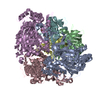 2xr8SC 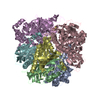 2xrxC 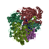 2xsoC C: citing same article ( S: Starting model for refinement |
|---|---|
| Similar structure data |


-Links
 PDBj
PDBj


- Assembly
| Deposited unit | 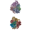
| ||||||||
|---|---|---|---|---|---|---|---|---|---|
| 1 | 
| ||||||||
| 2 | 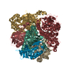
| ||||||||
| Unit cell |
|
-Components
-BIPHENYL DIOXYGENASE SUBUNIT ... , 2 types, 12 molecules ACEGIKBDFHJL
| #1: Protein | Mass: 51528.383 Da / Num. of mol.: 6 / Mutation: YES Source method: isolated from a genetically manipulated source Source: (gene. exp.) BURKHOLDERIA XENOVORANS (bacteria) / Strain: LB400 / Variant: P4 / Production host: #2: Protein | Mass: 22113.846 Da / Num. of mol.: 6 Source method: isolated from a genetically manipulated source Source: (gene. exp.) BURKHOLDERIA XENOVORANS (bacteria) / Strain: LB400 / Variant: P4 / Production host: |
|---|
-Non-polymers , 4 types, 1008 molecules 

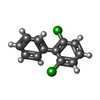

| #3: Chemical | ChemComp-FES /  Mass: 175.820 Da / Num. of mol.: 6 / Source method: obtained synthetically / Formula: Fe2S2 Mass: 175.820 Da / Num. of mol.: 6 / Source method: obtained synthetically / Formula: Fe2S2#4: Chemical | ChemComp-FE2 /  Mass: 55.845 Da / Num. of mol.: 6 / Source method: obtained synthetically / Formula: Fe Mass: 55.845 Da / Num. of mol.: 6 / Source method: obtained synthetically / Formula: Fe#5: Chemical |  Mass: 223.098 Da / Num. of mol.: 3 / Source method: obtained synthetically / Formula: C12H8Cl2 Mass: 223.098 Da / Num. of mol.: 3 / Source method: obtained synthetically / Formula: C12H8Cl2#6: Water | ChemComp-HOH / | Mass: 18.015 Da / Num. of mol.: 993 / Source method: isolated from a natural source / Formula: H2O |
|---|
-Details
| Compound details | ENGINEERED RESIDUE IN CHAIN A, THR 335 TO ALA ENGINEERED RESIDUE IN CHAIN C, THR 335 TO ALA ...ENGINEERED |
|---|
-Experimental details
-Experiment
| Experiment | Method: X-RAY DIFFRACTION / Number of used crystals: 1 |
|---|
- Sample preparation
| Crystal | Density Matthews: 2.3 Å3/Da / Density % sol: 47 % / Description: NONE |
|---|---|
| Crystal grow | pH: 6 / Details: PEG 8K, PIPES PH 6 |
-Data collection
| Diffraction | Mean temperature: 100 K |
|---|---|
| Diffraction source | Source: SYNCHROTRON / Site: APS  / Beamline: 22-ID / Wavelength: 1 / Beamline: 22-ID / Wavelength: 1 |
| Detector | Type: MARRESEARCH / Detector: CCD |
| Radiation | Protocol: SINGLE WAVELENGTH / Monochromatic (M) / Laue (L): M / Scattering type: x-ray |
| Radiation wavelength | Wavelength: 1 Å / Relative weight: 1 |
| Reflection | Resolution: 2.28→125 Å / Num. obs: 177300 / % possible obs: 86 % / Observed criterion σ(I): 1.5 / Redundancy: 4 % / Biso Wilson estimate: 47.5 Å2 / Rmerge(I) obs: 0.08 / Net I/σ(I): 18.6 |
| Reflection shell | Resolution: 2.28→2.38 Å / Redundancy: 1.2 % / Rmerge(I) obs: 0.35 / Mean I/σ(I) obs: 1.5 / % possible all: 51.4 |
- Processing
| Software |
| ||||||||||||||||||||||||||||||||||||||||||||||||||||||||||||||||||||||||||||||||||||||||||||||||||||||||||||||||||||||||||||||||||||||||||||||||||||||||||||||||||||||||||||||||||||||
|---|---|---|---|---|---|---|---|---|---|---|---|---|---|---|---|---|---|---|---|---|---|---|---|---|---|---|---|---|---|---|---|---|---|---|---|---|---|---|---|---|---|---|---|---|---|---|---|---|---|---|---|---|---|---|---|---|---|---|---|---|---|---|---|---|---|---|---|---|---|---|---|---|---|---|---|---|---|---|---|---|---|---|---|---|---|---|---|---|---|---|---|---|---|---|---|---|---|---|---|---|---|---|---|---|---|---|---|---|---|---|---|---|---|---|---|---|---|---|---|---|---|---|---|---|---|---|---|---|---|---|---|---|---|---|---|---|---|---|---|---|---|---|---|---|---|---|---|---|---|---|---|---|---|---|---|---|---|---|---|---|---|---|---|---|---|---|---|---|---|---|---|---|---|---|---|---|---|---|---|---|---|---|---|
| Refinement | Method to determine structure: MOLECULAR REPLACEMENT Starting model: PDB ENTRY 2XR8 Resolution: 2.29→138.68 Å / Cor.coef. Fo:Fc: 0.957 / Cor.coef. Fo:Fc free: 0.926 / SU B: 15.517 / SU ML: 0.194 / Cross valid method: THROUGHOUT / ESU R: 0.53 / ESU R Free: 0.273 / Stereochemistry target values: MAXIMUM LIKELIHOOD Details: HYDROGENS HAVE BEEN ADDED IN THE RIDING POSITIONS. ATOM RECORD CONTAINS SUM OF TLS AND RESIDUAL B FACTORS. ANISOU RECORD CONTAINS SUM OF TLS AND RESIDUAL U FACTORS.
| ||||||||||||||||||||||||||||||||||||||||||||||||||||||||||||||||||||||||||||||||||||||||||||||||||||||||||||||||||||||||||||||||||||||||||||||||||||||||||||||||||||||||||||||||||||||
| Solvent computation | Ion probe radii: 0.8 Å / Shrinkage radii: 0.8 Å / VDW probe radii: 1.2 Å / Solvent model: MASK | ||||||||||||||||||||||||||||||||||||||||||||||||||||||||||||||||||||||||||||||||||||||||||||||||||||||||||||||||||||||||||||||||||||||||||||||||||||||||||||||||||||||||||||||||||||||
| Displacement parameters | Biso mean: 50.381 Å2
| ||||||||||||||||||||||||||||||||||||||||||||||||||||||||||||||||||||||||||||||||||||||||||||||||||||||||||||||||||||||||||||||||||||||||||||||||||||||||||||||||||||||||||||||||||||||
| Refinement step | Cycle: LAST / Resolution: 2.29→138.68 Å
| ||||||||||||||||||||||||||||||||||||||||||||||||||||||||||||||||||||||||||||||||||||||||||||||||||||||||||||||||||||||||||||||||||||||||||||||||||||||||||||||||||||||||||||||||||||||
| Refine LS restraints |
|