 Movie
Movie Controller

[English] 日本語
 Yorodumi
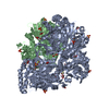
- PDB-1eg9: NAPHTHALENE 1,2-DIOXYGENASE WITH INDOLE BOUND IN THE ACTIVE SITE. -
+ Open data

- Basic information
| Entry | Database: PDB / ID: 1eg9 | ||||||
|---|---|---|---|---|---|---|---|
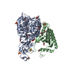
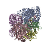
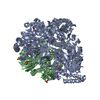
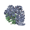
| Title | NAPHTHALENE 1,2-DIOXYGENASE WITH INDOLE BOUND IN THE ACTIVE SITE. | ||||||
 Components Components | (PROTEIN (NAPHTHALENE 1,2-DIOXYGENASE ...) x 2 | ||||||
 Keywords Keywords | OXIDOREDUCTASE / NON-HEME IRON DIOXYGENASE / ENZYME-SUBSTRATE COMPLEX | ||||||
| Function / homology |  Function and homology information Function and homology informationnaphthalene 1,2-dioxygenase / naphthalene 1,2-dioxygenase activity / 3-phenylpropionate catabolic process / dioxygenase activity / 2 iron, 2 sulfur cluster binding / iron ion binding Similarity search - Function | ||||||
| Biological species |  Pseudomonas putida (bacteria) Pseudomonas putida (bacteria) | ||||||
| Method |  X-RAY DIFFRACTION / SYNCHROTRON / Resolution: 1.6 Å X-RAY DIFFRACTION / SYNCHROTRON / Resolution: 1.6 Å | ||||||
 Authors Authors | Carredano, E. / Karlsson, A. / Kauppi, B. / Choudhury, D. / Parales, R.E. / Parales, J.V. / Lee, K. / Gibson, D.T. / Eklund, H. / Ramaswamy, S. | ||||||
 Citation Citation | Journal: J.Mol.Biol. / Year: 2000 Title: Substrate binding site of naphthalene 1,2-dioxygenase: functional implications of indole binding. Authors: Carredano, E. / Karlsson, A. / Kauppi, B. / Choudhury, D. / Parales, R.E. / Parales, J.V. / Lee, K. / Gibson, D.T. / Eklund, H. / Ramaswamy, S. | ||||||
| History |
|
- Structure visualization
| Structure viewer | Molecule: MolmilJmol/JSmol |
|---|
- Downloads & links
-Download
| PDBx/mmCIF format | 1eg9.cif.gz | 155.4 KB | Display | PDBx/mmCIF format |
|---|---|---|---|---|
| PDB format | pdb1eg9.ent.gz | 120.1 KB | Display | PDB format |
| PDBx/mmJSON format | 1eg9.json.gz | Tree view | PDBx/mmJSON format | |
| Others |  Other downloads Other downloads |
-Validation report
| Arichive directory | https://data.pdbj.org/pub/pdb/validation_reports/eg/1eg9ftp://data.pdbj.org/pub/pdb/validation_reports/eg/1eg9 | HTTPS FTP |
|---|
-Related structure data
| Similar structure data |
|---|


-Links
 PDBj
PDBj


- Assembly
| Deposited unit | 
| |||||||||
|---|---|---|---|---|---|---|---|---|---|---|
| 1 | 
| |||||||||
| 2 | x 6
| |||||||||
| Unit cell |
| |||||||||
| Components on special symmetry positions |
| |||||||||
| Details | THE ACTIVE ENZYME IS A ALPHA3BETA3 HEXAMER GENERATED BY THE THREEFOLD. |
-Components
-PROTEIN (NAPHTHALENE 1,2-DIOXYGENASE ... , 2 types, 2 molecules AB
| #1: Protein | Mass: 49664.355 Da / Num. of mol.: 1 Source method: isolated from a genetically manipulated source Source: (gene. exp.) Pseudomonas putida (bacteria) / Plasmid: PDTG14 / Production host: |
|---|---|
| #2: Protein | Mass: 22969.088 Da / Num. of mol.: 1 Source method: isolated from a genetically manipulated source Source: (gene. exp.) Pseudomonas putida (bacteria) / Plasmid: PDTG14 / Production host: |
-Non-polymers , 5 types, 618 molecules 


| #3: Chemical |  Mass: 96.063 Da / Num. of mol.: 3 / Source method: obtained synthetically / Formula: SO4 Mass: 96.063 Da / Num. of mol.: 3 / Source method: obtained synthetically / Formula: SO4#4: Chemical | ChemComp-FE / |  Mass: 55.845 Da / Num. of mol.: 1 / Source method: obtained synthetically / Formula: Fe Mass: 55.845 Da / Num. of mol.: 1 / Source method: obtained synthetically / Formula: Fe#5: Chemical | ChemComp-FES / |  Mass: 175.820 Da / Num. of mol.: 1 / Source method: obtained synthetically / Formula: Fe2S2 Mass: 175.820 Da / Num. of mol.: 1 / Source method: obtained synthetically / Formula: Fe2S2#6: Chemical | ChemComp-IND / |  Mass: 117.148 Da / Num. of mol.: 1 / Source method: obtained synthetically / Formula: C8H7N Mass: 117.148 Da / Num. of mol.: 1 / Source method: obtained synthetically / Formula: C8H7N#7: Water | ChemComp-HOH / | Mass: 18.015 Da / Num. of mol.: 612 / Source method: isolated from a natural source / Formula: H2O |
|---|
-Experimental details
-Experiment
| Experiment | Method: X-RAY DIFFRACTION / Number of used crystals: 1 |
|---|
- Sample preparation
| Crystal | Density Matthews: 2.72 Å3/Da / Density % sol: 54.82 % | |||||||||||||||||||||||||
|---|---|---|---|---|---|---|---|---|---|---|---|---|---|---|---|---|---|---|---|---|---|---|---|---|---|---|
| Crystal grow | pH: 6 Details: AMMONIUM SULPHATE 2M, MES 0.1M, DIOXANE 2-3%, pH 6.00 | |||||||||||||||||||||||||
| Crystal grow | *PLUS Temperature: 8 ℃ / Method: vapor diffusionDetails: drop consists of equal volume of protein and reservoir solutions | |||||||||||||||||||||||||
| Components of the solutions | *PLUS
|
-Data collection
| Diffraction | Mean temperature: 100 K |
|---|---|
| Diffraction source | Source: SYNCHROTRON / Site: ESRF  / Beamline: ID14-4 / Wavelength: 0.93 / Beamline: ID14-4 / Wavelength: 0.93 |
| Detector | Type: ADSC QUANTUM 4r / Detector: CCD / Date: Dec 5, 1998 |
| Radiation | Protocol: SINGLE WAVELENGTH / Monochromatic (M) / Laue (L): M / Scattering type: x-ray |
| Radiation wavelength | Wavelength: 0.93 Å / Relative weight: 1 |
| Reflection | Resolution: 1.6→20 Å / Num. obs: 896312 / % possible obs: 99.9 % / Redundancy: 8.8 % / Biso Wilson estimate: 16.8 Å2 / Rmerge(I) obs: 0.072 / Net I/σ(I): 11.3 |
| Reflection shell | Resolution: 1.6→20 Å / Redundancy: 4.9 % / Rmerge(I) obs: 0.307 / % possible all: 99 |
| Reflection | *PLUS Num. obs: 102843 / Num. measured all: 896312 |
| Reflection shell | *PLUS Lowest resolution: 1.63 Å / % possible obs: 99 % / Mean I/σ(I) obs: 3.31 |
- Processing
| Software |
| ||||||||||||
|---|---|---|---|---|---|---|---|---|---|---|---|---|---|
| Refinement | Resolution: 1.6→20 Å
| ||||||||||||
| Refinement step | Cycle: LAST / Resolution: 1.6→20 Å
| ||||||||||||
| Software | *PLUS Name: REFMAC / Classification: refinement | ||||||||||||
| Refine LS restraints | *PLUS
|
