 Movie
Movie Controller

[English] 日本語
 Yorodumi
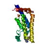
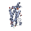
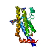
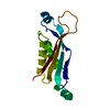
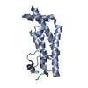
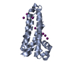
- PDB-1wq2: Neutron Crystal Structure Of Dissimilatory Sulfite Reductase D (DsrD) -
+ Open data

- Basic information
| Entry | Database: PDB / ID: 1wq2 | |||||||||
|---|---|---|---|---|---|---|---|---|---|---|
| Title | Neutron Crystal Structure Of Dissimilatory Sulfite Reductase D (DsrD) | |||||||||
 Components Components | Protein dsvD | |||||||||
 Keywords Keywords | UNKNOWN FUNCTION / neutron hydrogen hydration protein | |||||||||
| Function / homology |  Function and homology information Function and homology informationDissimilatory sulphite reductase D / Dissimilatory sulfite reductase D (DsrD) / Winged helix-like DNA-binding domain superfamily/Winged helix DNA-binding domain / Arc Repressor Mutant, subunit A / Winged helix DNA-binding domain superfamily / Winged helix-like DNA-binding domain superfamily / Orthogonal Bundle / Mainly Alpha Similarity search - Domain/homology | |||||||||
| Biological species |  Desulfovibrio vulgaris (bacteria) Desulfovibrio vulgaris (bacteria) | |||||||||
| Method | NEUTRON DIFFRACTION /  MOLECULAR REPLACEMENT / Resolution: 2.4 Å MOLECULAR REPLACEMENT / Resolution: 2.4 Å | |||||||||
 Authors Authors | Chatake, T. / Mizuno, N. / Voordouw, G. / Higuchi, Y. / Arai, S. / Tanaka, I. / Niimura, N. | |||||||||
 Citation Citation | Journal: Acta Crystallogr.,Sect.D / Year: 2003 Title: Crystallization and preliminary neutron analysis of the dissimilatory sulfite reductase D (DsrD) protein from the sulfate-reducing bacterium Desulfovibrio vulgaris. Authors: Chatake, T. / Mizuno, N. / Voordouw, G. / Higuchi, Y. / Arai, S. / Tanaka, I. / Niimura, N. #1: Journal: J.Synchrotron Radiat. / Year: 2004 Title: Hydration structures in proteins and neutron diffraction experiment on dissimilatory sul te reductase D (DsrD) Authors: Chatake, T. / Ostermann, A. / Kurihara, K. / Parak, F.G. / Mizuno, N. / Voordouw, G. / Higuchi, Y. / Tanaka, I. / Niimura, N. | |||||||||
| History |
|
- Structure visualization
| Structure viewer | Molecule: MolmilJmol/JSmol |
|---|
- Downloads & links
-Download
| PDBx/mmCIF format | 1wq2.cif.gz | 62 KB | Display | PDBx/mmCIF format |
|---|---|---|---|---|
| PDB format | pdb1wq2.ent.gz | 46.4 KB | Display | PDB format |
| PDBx/mmJSON format | 1wq2.json.gz | Tree view | PDBx/mmJSON format | |
| Others |  Other downloads Other downloads |
-Validation report
| Summary document | 1wq2_validation.pdf.gz | 355.5 KB | Display | wwPDB validaton report |
|---|---|---|---|---|
| Full document | 1wq2_full_validation.pdf.gz | 355.5 KB | Display | |
| Data in XML | 1wq2_validation.xml.gz | 4.2 KB | Display | |
| Data in CIF | 1wq2_validation.cif.gz | 6.9 KB | Display | |
| Arichive directory | https://data.pdbj.org/pub/pdb/validation_reports/wq/1wq2ftp://data.pdbj.org/pub/pdb/validation_reports/wq/1wq2 | HTTPS FTP |
-Related structure data
| Related structure data | 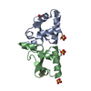 1ucrS S: Starting model for refinement |
|---|---|
| Similar structure data |


-Links
 PDBj
PDBj

- Assembly
| Deposited unit | 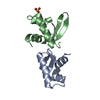
| ||||||||
|---|---|---|---|---|---|---|---|---|---|
| 1 |
| ||||||||
| Unit cell |
|
-Components
| #1: Protein | Mass: 8841.918 Da / Num. of mol.: 2 Source method: isolated from a genetically manipulated source Source: (gene. exp.) Desulfovibrio vulgaris (bacteria) / Production host: #2: Chemical | ChemComp-SO4 / |   Mass: 96.063 Da / Num. of mol.: 1 / Source method: obtained synthetically / Formula: SO4 Mass: 96.063 Da / Num. of mol.: 1 / Source method: obtained synthetically / Formula: SO4#3: Chemical | ChemComp-DOD / |   Mass: 18.015 Da / Num. of mol.: 99 / Source method: isolated from a natural source / Formula: D2O Mass: 18.015 Da / Num. of mol.: 99 / Source method: isolated from a natural source / Formula: D2O |
|---|
-Experimental details
-Experiment
| Experiment | Method: NEUTRON DIFFRACTION / Number of used crystals: 1 |
|---|
- Sample preparation
| Crystal grow | Temperature: 293 K / Method: vapor diffusion, sitting drop / pH: 5.3 Details: deuterated ammoniu sulfate, deuterated Tris-HCl, pH 5.3, VAPOR DIFFUSION, SITTING DROP, temperature 293K |
|---|
-Data collection
| Diffraction | Mean temperature: 293 K |
|---|---|
| Diffraction source | Wavelength: 2.88 Å |
| Detector | Detector: IMAGE PLATE / Date: May 5, 2003 / Details: monochromator BIX-3 |
| Radiation | Monochromator: ellastically bent silicon / Protocol: SINGLE WAVELENGTH / Monochromatic (M) / Laue (L): M / Scattering type: x-ray |
| Radiation wavelength | Wavelength: 2.88 Å / Relative weight: 1 |
| Reflection | Resolution: 2.4→100 Å / Num. all: 7572 / Num. obs: 7001 / % possible obs: 92.5 % / Observed criterion σ(F): 0 / Observed criterion σ(I): 1 / Rmerge(I) obs: 0.143 |
| Reflection shell | Resolution: 2.4→2.49 Å / Rmerge(I) obs: 0.395 / Mean I/σ(I) obs: 2.3 / Num. unique all: 534 / % possible all: 82.1 |
- Processing
| Software |
| |||||||||||||||||||||||||
|---|---|---|---|---|---|---|---|---|---|---|---|---|---|---|---|---|---|---|---|---|---|---|---|---|---|---|
| Refinement | Method to determine structure: MOLECULAR REPLACEMENT Starting model: PDB ENTRY 1UCR Resolution: 2.4→20 Å / Isotropic thermal model: Isotropic / Cross valid method: THROUGHOUT / σ(F): 1 / Stereochemistry target values: Engh & Huber
| |||||||||||||||||||||||||
| Displacement parameters | Biso mean: 18.3 Å2 | |||||||||||||||||||||||||
| Refine analyze |
| |||||||||||||||||||||||||
| Refinement step | Cycle: LAST / Resolution: 2.4→20 Å
| |||||||||||||||||||||||||
| Refine LS restraints |
|
