 Movie
Movie Controller

[English] 日本語
 Yorodumi
- PDB-1wox: Crystal structure of heme oxygenase-2 from Synechocystis sp. PCC ... -
+ Open data

- Basic information
| Entry | Database: PDB / ID: 1wox | ||||||
|---|---|---|---|---|---|---|---|
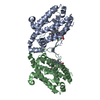
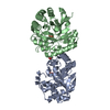
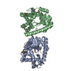
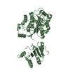
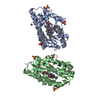
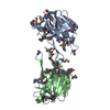
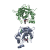
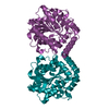
| Title | Crystal structure of heme oxygenase-2 from Synechocystis sp. PCC 6803 in complex with heme and NO | ||||||
 Components Components | Heme oxygenase 2 | ||||||
 Keywords Keywords | OXIDOREDUCTASE / HOMO-DIMER / NO-bound heme complex | ||||||
| Function / homology |  Function and homology information Function and homology informationheme oxygenase (biliverdin-producing) / heme oxidation / heme oxygenase (decyclizing) activity / heme catabolic process / photosynthesis / response to oxidative stress / heme binding / metal ion binding / plasma membrane Similarity search - Function | ||||||
| Biological species |  | ||||||
| Method |  X-RAY DIFFRACTION / SYNCHROTRON / MOLECULAR REPLACEMENT / Resolution: 2.1 Å X-RAY DIFFRACTION / SYNCHROTRON / MOLECULAR REPLACEMENT / Resolution: 2.1 Å | ||||||
 Authors Authors | Sugishima, M. / Hagiwara, Y. / Zhang, X. / Yoshida, T. / Migita, C.T. / Fukuyama, K. | ||||||
 Citation Citation | Journal: Biochemistry / Year: 2005 Title: Crystal structure of dimeric heme oxygenase-2 from Synechocystis sp. PCC 6803 in complex with heme. Authors: Sugishima, M. / Hagiwara, Y. / Zhang, X. / Yoshida, T. / Migita, C.T. / Fukuyama, K. | ||||||
| History |
|
- Structure visualization
| Structure viewer | Molecule: MolmilJmol/JSmol |
|---|
- Downloads & links
-Download
| PDBx/mmCIF format | 1wox.cif.gz | 118.3 KB | Display | PDBx/mmCIF format |
|---|---|---|---|---|
| PDB format | pdb1wox.ent.gz | 90.8 KB | Display | PDB format |
| PDBx/mmJSON format | 1wox.json.gz | Tree view | PDBx/mmJSON format | |
| Others |  Other downloads Other downloads |
-Validation report
| Arichive directory | https://data.pdbj.org/pub/pdb/validation_reports/wo/1woxftp://data.pdbj.org/pub/pdb/validation_reports/wo/1wox | HTTPS FTP |
|---|
-Related structure data
| Related structure data |  1wovC  1wowC 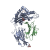 1w0vS C: citing same article ( S: Starting model for refinement |
|---|---|
| Similar structure data |


-Links
 PDBj
PDBj


- Assembly
| Deposited unit | 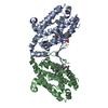
| ||||||||
|---|---|---|---|---|---|---|---|---|---|
| 1 |
| ||||||||
| Unit cell |
| ||||||||
| Details | The biological assembly is a dimer in the asymmmetric unit. |
-Components
| #1: Protein | Mass: 28572.312 Da / Num. of mol.: 2 Source method: isolated from a genetically manipulated source Source: (gene. exp.) References: UniProt: P74133, heme oxygenase (biliverdin-producing) #2: Chemical |   Mass: 616.487 Da / Num. of mol.: 2 / Source method: obtained synthetically / Formula: C34H32FeN4O4 Mass: 616.487 Da / Num. of mol.: 2 / Source method: obtained synthetically / Formula: C34H32FeN4O4#3: Chemical |   Mass: 30.006 Da / Num. of mol.: 2 / Source method: obtained synthetically / Formula: NO Mass: 30.006 Da / Num. of mol.: 2 / Source method: obtained synthetically / Formula: NO#4: Water | ChemComp-HOH / |  Mass: 18.015 Da / Num. of mol.: 311 / Source method: isolated from a natural source / Formula: H2O Mass: 18.015 Da / Num. of mol.: 311 / Source method: isolated from a natural source / Formula: H2O |
|---|
-Experimental details
-Experiment
| Experiment | Method: X-RAY DIFFRACTION / Number of used crystals: 1 |
|---|
- Sample preparation
| Crystal | Density Matthews: 2.5 Å3/Da / Density % sol: 50.8 % |
|---|---|
| Crystal grow | Temperature: 293 K / Method: vapor diffusion, hanging drop / pH: 7 Details: pottasium sodium tartrate, pottasium phosphate, pH 7.0, VAPOR DIFFUSION, HANGING DROP, temperature 293K |
-Data collection
| Diffraction | Mean temperature: 100 K |
|---|---|
| Diffraction source | Source: SYNCHROTRON / Site: SPring-8  / Beamline: BL41XU / Wavelength: 1.046 Å / Beamline: BL41XU / Wavelength: 1.046 Å |
| Detector | Type: MARRESEARCH / Detector: CCD / Date: May 29, 2004 |
| Radiation | Monochromator: Si(111) Double monochromator / Protocol: SINGLE WAVELENGTH / Monochromatic (M) / Laue (L): M / Scattering type: x-ray |
| Radiation wavelength | Wavelength: 1.046 Å / Relative weight: 1 |
| Reflection | Resolution: 2.1→50 Å / Num. all: 33206 / Num. obs: 33206 / % possible obs: 100 % / Observed criterion σ(F): 0 / Observed criterion σ(I): 0 |
| Reflection shell | Resolution: 2.1→2.21 Å / % possible all: 100 |
- Processing
| Software |
| ||||||||||||||||||||
|---|---|---|---|---|---|---|---|---|---|---|---|---|---|---|---|---|---|---|---|---|---|
| Refinement | Method to determine structure: MOLECULAR REPLACEMENT Starting model: PDB ENTRY 1W0V Resolution: 2.1→20 Å / Cross valid method: THROUGHOUT / σ(F): 0 / Stereochemistry target values: Engh & Huber
| ||||||||||||||||||||
| Refine analyze |
| ||||||||||||||||||||
| Refinement step | Cycle: LAST / Resolution: 2.1→20 Å
| ||||||||||||||||||||
| Refine LS restraints |
| ||||||||||||||||||||
| LS refinement shell | Resolution: 2.1→2.18 Å
|
