 Movie
Movie Controller


+ Open data

- Basic information
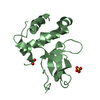
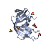
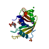
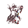
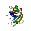
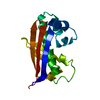
| Entry | Database: PDB / ID: 1wol | ||||||
|---|---|---|---|---|---|---|---|
| Title | Crystal Structure of ST0689, an archaeal HEPN homologue | ||||||
 Components Components | 122aa long conserved hypothetical protein | ||||||
 Keywords Keywords | UNKNOWN FUNCTION / ALPHA HELIX / LOOP | ||||||
| Function / homology |  Function and homology information Function and homology informationHEPN domain profile. / Higher Eukarytoes and Prokaryotes Nucleotide-binding domain / HEPN domain / HEPN domain / Nucleotidyltransferases domain 2 / Four Helix Bundle (Hemerythrin (Met), subunit A) / Up-down Bundle / Mainly Alpha Similarity search - Domain/homology | ||||||
| Biological species |   Sulfolobus tokodaii (archaea) Sulfolobus tokodaii (archaea) | ||||||
| Method |  X-RAY DIFFRACTION / SYNCHROTRON / MAD / Resolution: 1.62 Å X-RAY DIFFRACTION / SYNCHROTRON / MAD / Resolution: 1.62 Å | ||||||
 Authors Authors | Arakawa, T. / Akutsu, J. / Yohda, M. | ||||||
 Citation Citation | Journal: To be Published Title: Crystal Structure Analysis of ST0689, an archaeal HEPN protein Authors: Arakawa, T. / Akutsu, J. / Yohda, M. | ||||||
| History |
|
- Structure visualization
| Structure viewer | Molecule: MolmilJmol/JSmol |
|---|
- Downloads & links
-Download
| PDBx/mmCIF format | 1wol.cif.gz | 40.2 KB | Display | PDBx/mmCIF format |
|---|---|---|---|---|
| PDB format | pdb1wol.ent.gz | 27.6 KB | Display | PDB format |
| PDBx/mmJSON format | 1wol.json.gz | Tree view | PDBx/mmJSON format | |
| Others |  Other downloads Other downloads |
-Validation report
| Arichive directory | https://data.pdbj.org/pub/pdb/validation_reports/wo/1wolftp://data.pdbj.org/pub/pdb/validation_reports/wo/1wol | HTTPS FTP |
|---|
-Related structure data
| Similar structure data |
|---|


-Links
 PDBj
PDBj- Assembly
| Deposited unit | 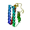
| ||||||||
|---|---|---|---|---|---|---|---|---|---|
| 1 |
| ||||||||
| Unit cell |
| ||||||||
| Details | The biological assembly seems to be a monomer |
-Components
| #1: Protein | Mass: 14183.036 Da / Num. of mol.: 1 Source method: isolated from a genetically manipulated source Source: (gene. exp.) Sulfolobus tokodaii (archaea) / Strain: str. 7 / Plasmid: pET23a / Production host:  | ||||
|---|---|---|---|---|---|
| #2: Chemical | 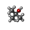  Mass: 74.122 Da / Num. of mol.: 2 / Source method: obtained synthetically / Formula: C4H10O Mass: 74.122 Da / Num. of mol.: 2 / Source method: obtained synthetically / Formula: C4H10O#3: Water | ChemComp-HOH / |  Mass: 18.015 Da / Num. of mol.: 84 / Source method: isolated from a natural source / Formula: H2O Mass: 18.015 Da / Num. of mol.: 84 / Source method: isolated from a natural source / Formula: H2OHas protein modification | Y | |
-Experimental details
-Experiment
| Experiment | Method: X-RAY DIFFRACTION |
|---|
- Sample preparation
| Crystal | Density Matthews: 2.5 Å3/Da / Density % sol: 50 % |
|---|---|
| Crystal grow | Temperature: 293 K / Method: vapor diffusion, hanging drop / pH: 4.8 Details: tert-butanol, citrate, pH 4.8, VAPOR DIFFUSION, HANGING DROP, temperature 293K |
-Data collection
| Diffraction | Mean temperature: 100 K | ||||||||||||
|---|---|---|---|---|---|---|---|---|---|---|---|---|---|
| Diffraction source | Source: SYNCHROTRON / Site: SPring-8  / Beamline: BL38B1 / Wavelength: 0.9765, 0.97894, 0.97924 / Beamline: BL38B1 / Wavelength: 0.9765, 0.97894, 0.97924 | ||||||||||||
| Detector | Type: ADSC QUANTUM 4 / Detector: CCD / Date: Jul 2, 2004 | ||||||||||||
| Radiation | Monochromator: GRAPHITE / Protocol: MAD / Monochromatic (M) / Laue (L): M / Scattering type: x-ray | ||||||||||||
| Radiation wavelength |
| ||||||||||||
| Reflection | Resolution: 1.62→50 Å / Biso Wilson estimate: 22.2 Å2 | ||||||||||||
| Reflection shell | Resolution: 1.62→1.68 Å / Rsym value: 0.177 / % possible all: 100 |
- Processing
| Software |
| ||||||||||||||||||||||||||||||||||||
|---|---|---|---|---|---|---|---|---|---|---|---|---|---|---|---|---|---|---|---|---|---|---|---|---|---|---|---|---|---|---|---|---|---|---|---|---|---|
| Refinement | Method to determine structure: MAD / Resolution: 1.62→41.35 Å / Rfactor Rfree error: 0.004 / Data cutoff high absF: 335259.04 / Data cutoff low absF: 0 / Isotropic thermal model: RESTRAINED / Cross valid method: THROUGHOUT / σ(F): 0 / Stereochemistry target values: Engh & Huber
| ||||||||||||||||||||||||||||||||||||
| Solvent computation | Solvent model: FLAT MODEL / Bsol: 41.7591 Å2 / ksol: 0.378148 e/Å3 | ||||||||||||||||||||||||||||||||||||
| Displacement parameters | Biso mean: 27 Å2
| ||||||||||||||||||||||||||||||||||||
| Refine analyze |
| ||||||||||||||||||||||||||||||||||||
| Refinement step | Cycle: LAST / Resolution: 1.62→41.35 Å
| ||||||||||||||||||||||||||||||||||||
| Refine LS restraints |
| ||||||||||||||||||||||||||||||||||||
| LS refinement shell | Resolution: 1.62→1.72 Å / Rfactor Rfree error: 0.011 / Total num. of bins used: 6
| ||||||||||||||||||||||||||||||||||||
| Xplor file |
|
