Entry Database : PDB / ID : 1wokTitle Crystal structure of catalytic domain of human poly(ADP-ribose) polymerase complexed with a quinoxaline-type inhibitor Poly [ADP-ribose] polymerase-1 Keywords / Function / homology Function Domain/homology Component
/ / / / / / / / / / / / / / / / / / / / / / / / / / / / / / / / / / / / / / / / / / / / / / / / / / / / / / / / / / / / / / / / / / / / / / / / / / / / / / / / / / / / / / / / / / / / / / / / / / / / / / / / / / / / / / / / / / / / / / / / / / / / / / / / / / / / / / / / / / / / / / / / / / / / / / / / / / / / Biological species Homo sapiens (human)Method / / / Resolution : 3 Å Authors Iwashita, A. / Hattori, K. / Yamamoto, H. / Ishida, J. / Kido, Y. / Kamijo, K. / Murano, K. / Miyake, H. / Kinoshita, T. / Warizaya, M. ...Iwashita, A. / Hattori, K. / Yamamoto, H. / Ishida, J. / Kido, Y. / Kamijo, K. / Murano, K. / Miyake, H. / Kinoshita, T. / Warizaya, M. / Ohkubo, M. / Matsuoka, N. / Mutoh, S. Journal : Febs Lett. / Year : 2005Title : Discovery of quinazolinone and quinoxaline derivatives as potent and selective poly(ADP-ribose) polymerase-1/2 inhibitors.Authors : Iwashita, A. / Hattori, K. / Yamamoto, H. / Ishida, J. / Kido, Y. / Kamijo, K. / Murano, K. / Miyake, H. / Kinoshita, T. / Warizaya, M. / Ohkubo, M. / Matsuoka, N. / Mutoh, S. History Deposition Aug 20, 2004 Deposition site / Processing site Revision 1.0 Mar 15, 2005 Provider / Type Revision 1.1 Apr 30, 2008 Group Revision 1.2 Jul 13, 2011 Group Revision 1.3 Oct 11, 2017 Group / Category Revision 1.4 Oct 25, 2023 Group Data collection / Database references ... Data collection / Database references / Derived calculations / Refinement description Category chem_comp_atom / chem_comp_bond ... chem_comp_atom / chem_comp_bond / database_2 / pdbx_initial_refinement_model / struct_site Item _database_2.pdbx_DOI / _database_2.pdbx_database_accession ... _database_2.pdbx_DOI / _database_2.pdbx_database_accession / _struct_site.pdbx_auth_asym_id / _struct_site.pdbx_auth_comp_id / _struct_site.pdbx_auth_seq_id
Show all Show less
 Movie
Movie Controller

 Yorodumi
 Open data
 Basic information
 Components
 Keywords
 Function and homology information
 Homo sapiens (human)
 X-RAY DIFFRACTION /
  Authors
 Citation
 Structure visualization
 Downloads & links
 Other downloads
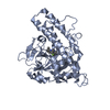

 PDBj
PDBj


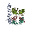
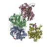
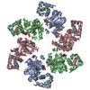
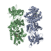
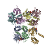
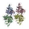
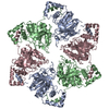
 Assembly
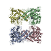

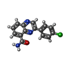
 Mass: 283.712 Da / Num. of mol.: 4 / Source method: obtained synthetically / Formula: C15H10ClN3O
 Sample preparation
 / Beamline: BL26B2 / Wavelength: 1 Å
 Processing