 Movie
Movie Controller

[English] 日本語
 Yorodumi
- PDB-1wlz: Crystal structure of DJBP fragment which was obtained by limited ... -
+ Open data

- Basic information
| Entry | Database: PDB / ID: 1wlz | ||||||
|---|---|---|---|---|---|---|---|
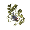
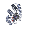
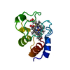
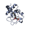
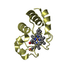
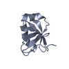
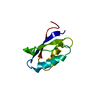
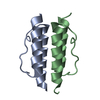
| Title | Crystal structure of DJBP fragment which was obtained by limited proteolysis | ||||||
 Components Components | CAP-binding protein complex interacting protein 1 isoform a | ||||||
 Keywords Keywords | UNKNOWN FUNCTION / EF-hand like | ||||||
| Function / homology |  Function and homology information Function and homology informationaxonemal A tubule inner sheath / flagellated sperm motility / sperm flagellum / calcium ion binding / nucleoplasm Similarity search - Function | ||||||
| Biological species |  Homo sapiens (human) Homo sapiens (human) | ||||||
| Method |  X-RAY DIFFRACTION / SYNCHROTRON / MAD, SIR / Resolution: 1.6 Å X-RAY DIFFRACTION / SYNCHROTRON / MAD, SIR / Resolution: 1.6 Å | ||||||
 Authors Authors | Honbou, K. / Suzuki, N. / Horiuchi, M. / Taira, T. / Niki, T. / Ariga, H. / Inagaki, F. | ||||||
 Citation Citation | Journal: To be Published Title: Crystal Structure of DJBP Fragment which was obtained by Limited Proteolysis Authors: Honbou, K. / Suzuki, N. / Horiuchi, M. / Taira, T. / Niki, T. / Ariga, H. / Inagaki, F. | ||||||
| History |
|
- Structure visualization
| Structure viewer | Molecule: MolmilJmol/JSmol |
|---|
- Downloads & links
-Download
| PDBx/mmCIF format | 1wlz.cif.gz | 90 KB | Display | PDBx/mmCIF format |
|---|---|---|---|---|
| PDB format | pdb1wlz.ent.gz | 67.4 KB | Display | PDB format |
| PDBx/mmJSON format | 1wlz.json.gz | Tree view | PDBx/mmJSON format | |
| Others |  Other downloads Other downloads |
-Validation report
| Summary document | 1wlz_validation.pdf.gz | 446.9 KB | Display | wwPDB validaton report |
|---|---|---|---|---|
| Full document | 1wlz_full_validation.pdf.gz | 449.8 KB | Display | |
| Data in XML | 1wlz_validation.xml.gz | 20 KB | Display | |
| Data in CIF | 1wlz_validation.cif.gz | 29.4 KB | Display | |
| Arichive directory | https://data.pdbj.org/pub/pdb/validation_reports/wl/1wlzftp://data.pdbj.org/pub/pdb/validation_reports/wl/1wlz | HTTPS FTP |
-Related structure data
| Similar structure data |
|---|


-Links
 PDBj
PDBj

- Assembly
| Deposited unit | 
| ||||||||
|---|---|---|---|---|---|---|---|---|---|
| 1 | 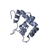
| ||||||||
| 2 | 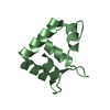
| ||||||||
| 3 | 
| ||||||||
| 4 | 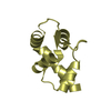
| ||||||||
| Unit cell |
|
-Components
| #1: Protein | Mass: 12062.537 Da / Num. of mol.: 4 / Fragment: RESIDUES 229-329 Source method: isolated from a genetically manipulated source Source: (gene. exp.) Homo sapiens (human) / Plasmid: pGEX-6P-1 / Species (production host): Escherichia coli / Production host:  #2: Water | ChemComp-HOH / |  Mass: 18.015 Da / Num. of mol.: 491 / Source method: isolated from a natural source / Formula: H2O Mass: 18.015 Da / Num. of mol.: 491 / Source method: isolated from a natural source / Formula: H2O |
|---|
-Experimental details
-Experiment
| Experiment | Method: X-RAY DIFFRACTION / Number of used crystals: 1 |
|---|
- Sample preparation
| Crystal | Density Matthews: 2.06 Å3/Da / Density % sol: 40.36 % |
|---|---|
| Crystal grow | Temperature: 293 K / Method: vapor diffusion, sitting drop / pH: 4.6 Details: 30% PEG MME 2000, 0.1M Na Acetate pH 4.6, 0.2M Ammonium Sulfate, VAPOR DIFFUSION, SITTING DROP, temperature 293K |
-Data collection
| Diffraction | Mean temperature: 100 K |
|---|---|
| Diffraction source | Source: SYNCHROTRON / Site: SPring-8  / Beamline: BL38B1 / Wavelength: 1 Å / Beamline: BL38B1 / Wavelength: 1 Å |
| Detector | Type: ADSC QUANTUM 4 / Detector: CCD / Date: Oct 30, 2002 |
| Radiation | Monochromator: SI / Protocol: SINGLE WAVELENGTH / Monochromatic (M) / Laue (L): M / Scattering type: x-ray |
| Radiation wavelength | Wavelength: 1 Å / Relative weight: 1 |
| Reflection | Resolution: 1.6→100 Å / Num. all: 51566 / Num. obs: 51566 / % possible obs: 99.4 % / Observed criterion σ(F): 0 / Observed criterion σ(I): -3 / Redundancy: 3.7 % / Biso Wilson estimate: 17.6 Å2 / Rmerge(I) obs: 0.042 |
| Reflection shell | Resolution: 1.6→1.66 Å / % possible all: 95.1 |
- Processing
| Software |
| ||||||||||||||||||||||||||||||||||||||||||||||||||||||||||||||||||||||||||||||||
|---|---|---|---|---|---|---|---|---|---|---|---|---|---|---|---|---|---|---|---|---|---|---|---|---|---|---|---|---|---|---|---|---|---|---|---|---|---|---|---|---|---|---|---|---|---|---|---|---|---|---|---|---|---|---|---|---|---|---|---|---|---|---|---|---|---|---|---|---|---|---|---|---|---|---|---|---|---|---|---|---|---|
| Refinement | Method to determine structure: MAD, SIR / Resolution: 1.6→19.95 Å / Rfactor Rfree error: 0.003 / Data cutoff high absF: 455686.28 / Data cutoff low absF: 0 / Isotropic thermal model: RESTRAINED / Cross valid method: THROUGHOUT / σ(F): 0 / Stereochemistry target values: Engh & Huber
| ||||||||||||||||||||||||||||||||||||||||||||||||||||||||||||||||||||||||||||||||
| Solvent computation | Solvent model: FLAT MODEL / Bsol: 49.872 Å2 / ksol: 0.377187 e/Å3 | ||||||||||||||||||||||||||||||||||||||||||||||||||||||||||||||||||||||||||||||||
| Displacement parameters | Biso mean: 17.7 Å2
| ||||||||||||||||||||||||||||||||||||||||||||||||||||||||||||||||||||||||||||||||
| Refine analyze |
| ||||||||||||||||||||||||||||||||||||||||||||||||||||||||||||||||||||||||||||||||
| Refinement step | Cycle: LAST / Resolution: 1.6→19.95 Å
| ||||||||||||||||||||||||||||||||||||||||||||||||||||||||||||||||||||||||||||||||
| Refine LS restraints |
| ||||||||||||||||||||||||||||||||||||||||||||||||||||||||||||||||||||||||||||||||
| LS refinement shell | Resolution: 1.6→1.7 Å / Rfactor Rfree error: 0.009 / Total num. of bins used: 6
| ||||||||||||||||||||||||||||||||||||||||||||||||||||||||||||||||||||||||||||||||
| Xplor file |
|
