 Movie
Movie Controller

[English] 日本語
 Yorodumi
- PDB-1wle: Crystal Structure of mammalian mitochondrial seryl-tRNA synthetas... -
+ Open data

- Basic information
| Entry | Database: PDB / ID: 1wle | ||||||
|---|---|---|---|---|---|---|---|
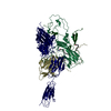
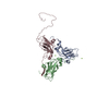
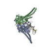
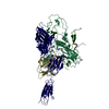
| Title | Crystal Structure of mammalian mitochondrial seryl-tRNA synthetase complexed with seryl-adenylate | ||||||
 Components Components | Seryl-tRNA synthetase | ||||||
 Keywords Keywords | LIGASE | ||||||
| Function / homology |  Function and homology information Function and homology informationmitochondrial seryl-tRNA aminoacylation / serine-tRNA ligase / serine-tRNA ligase activity / seryl-tRNA aminoacylation / tRNA binding / mitochondrial matrix / mitochondrion / ATP binding Similarity search - Function | ||||||
| Biological species |  | ||||||
| Method |  X-RAY DIFFRACTION / SYNCHROTRON / MOLECULAR REPLACEMENT / Resolution: 1.65 Å X-RAY DIFFRACTION / SYNCHROTRON / MOLECULAR REPLACEMENT / Resolution: 1.65 Å | ||||||
 Authors Authors | Chimnaronk, S. / Jeppesen, M.G. / Suzuki, T. / Nyborg, J. / Watanabe, K. | ||||||
 Citation Citation | Journal: Embo J. / Year: 2005 Title: Dual-mode recognition of noncanonical tRNAs(Ser) by seryl-tRNA synthetase in mammalian mitochondria Authors: Chimnaronk, S. / Jeppesen, M.G. / Suzuki, T. / Nyborg, J. / Watanabe, K. #1: Journal: J.Biol.Chem. / Year: 2000 Title: Characterization and tRNA recognition of mammalian mitochondrial seryl-tRNA synthetase Authors: Yokogawa, T. / Shimada, N. / Takeuchi, N. / Benkowski, L. / Suzuki, T. / Omori, A. / Ueda, T. / Nishikawa, K. / Spremulli, L.L. / Watanabe, K. #2: Journal: J.Biol.Chem. / Year: 2001 Title: Dual mode recognition of two isoacceptor tRNAs by mammalian mitochondrial seryl-tRNA synthetase Authors: Shimada, N. / Suzuki, T. / Watanabe, K. #3: Journal: ACTA CRYSTALLOGR.,SECT.D / Year: 2004 Title: Crystallization and preliminary X-ray diffraction study of mammalian mitochondrial seryl-tRNA synthetase Authors: Chimnaronk, S. / Jeppesen, M.G. / Shimada, N. / Suzuki, T. / Nyborg, J. / Watanabe, K. | ||||||
| History |
|
- Structure visualization
| Structure viewer | Molecule: MolmilJmol/JSmol |
|---|
- Downloads & links
-Download
| PDBx/mmCIF format | 1wle.cif.gz | 209.1 KB | Display | PDBx/mmCIF format |
|---|---|---|---|---|
| PDB format | pdb1wle.ent.gz | 166.1 KB | Display | PDB format |
| PDBx/mmJSON format | 1wle.json.gz | Tree view | PDBx/mmJSON format | |
| Others |  Other downloads Other downloads |
-Validation report
| Arichive directory | https://data.pdbj.org/pub/pdb/validation_reports/wl/1wleftp://data.pdbj.org/pub/pdb/validation_reports/wl/1wle | HTTPS FTP |
|---|
-Related structure data
| Related structure data |  1sryS S: Starting model for refinement |
|---|---|
| Similar structure data |


-Links
 PDBj
PDBj
- Assembly
| Deposited unit | 
| ||||||||
|---|---|---|---|---|---|---|---|---|---|
| 1 |
| ||||||||
| Unit cell |
| ||||||||
| Details | The biological assembly is a homodimer found in the asymmetric unit. |
-Components
| #1: Protein | Mass: 56563.047 Da / Num. of mol.: 2 Source method: isolated from a genetically manipulated source Source: (gene. exp.)  #2: Chemical |   Mass: 434.299 Da / Num. of mol.: 2 / Source method: obtained synthetically / Formula: C13H19N6O9P Mass: 434.299 Da / Num. of mol.: 2 / Source method: obtained synthetically / Formula: C13H19N6O9P#3: Water | ChemComp-HOH / |  Mass: 18.015 Da / Num. of mol.: 611 / Source method: isolated from a natural source / Formula: H2O Mass: 18.015 Da / Num. of mol.: 611 / Source method: isolated from a natural source / Formula: H2O |
|---|
-Experimental details
-Experiment
| Experiment | Method: X-RAY DIFFRACTION / Number of used crystals: 1 |
|---|
- Sample preparation
| Crystal | Density Matthews: 2.8 Å3/Da / Density % sol: 55.1 % |
|---|---|
| Crystal grow | Temperature: 293 K / Method: vapor diffusion, sitting drop / pH: 5.5 Details: PEG 8000, lithium sulfate, MES-NaOH, DTT, pH 5.5, VAPOR DIFFUSION, SITTING DROP, temperature 293.0K |
-Data collection
| Diffraction | Mean temperature: 100 K |
|---|---|
| Diffraction source | Source: SYNCHROTRON / Site: EMBL/DESY, HAMBURG  / Beamline: X11 / Wavelength: 0.8123 Å / Beamline: X11 / Wavelength: 0.8123 Å |
| Detector | Type: MARRESEARCH / Detector: CCD / Date: Nov 6, 2003 / Details: Bent mirror |
| Radiation | Monochromator: Triangular / Protocol: SINGLE WAVELENGTH / Monochromatic (M) / Laue (L): M / Scattering type: x-ray |
| Radiation wavelength | Wavelength: 0.8123 Å / Relative weight: 1 |
| Reflection | Resolution: 1.65→99 Å / Num. all: 145408 / Num. obs: 145408 / % possible obs: 96.6 % / Observed criterion σ(F): 0 / Observed criterion σ(I): -0.3 / Redundancy: 6.3 % / Biso Wilson estimate: 21.206 Å2 / Rmerge(I) obs: 0.05 / Net I/σ(I): 30.7 |
| Reflection shell | Resolution: 1.65→1.69 Å / Redundancy: 4.3 % / Rmerge(I) obs: 0.372 / Mean I/σ(I) obs: 1.8 / Num. unique all: 9594 / % possible all: 96.9 |
- Processing
| Software |
| |||||||||||||||||||||||||||
|---|---|---|---|---|---|---|---|---|---|---|---|---|---|---|---|---|---|---|---|---|---|---|---|---|---|---|---|---|
| Refinement | Method to determine structure: MOLECULAR REPLACEMENT Starting model: PDB Entry 1SRY Resolution: 1.65→10 Å / Isotropic thermal model: isotropic / Cross valid method: THROUGHOUT / σ(F): 0 / Stereochemistry target values: Engh & Huber
| |||||||||||||||||||||||||||
| Solvent computation | Solvent model: THROUGHOUT / Bsol: 63.3399 Å2 / ksol: 0.483133 e/Å3 | |||||||||||||||||||||||||||
| Displacement parameters | Biso mean: 28.5261 Å2
| |||||||||||||||||||||||||||
| Refine analyze |
| |||||||||||||||||||||||||||
| Refinement step | Cycle: LAST / Resolution: 1.65→10 Å
| |||||||||||||||||||||||||||
| Refine LS restraints |
| |||||||||||||||||||||||||||
| LS refinement shell | Resolution: 1.64→1.7 Å / Total num. of bins used: 10
| |||||||||||||||||||||||||||
| Xplor file | Serial no: 1 / Param file: SMP.PARAM / Topol file: SMP.TOP |
