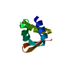
 Movie
Movie Controller


+ Open data

- Basic information
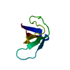
| Entry | Database: PDB / ID: 1wdx | ||||||
|---|---|---|---|---|---|---|---|
| Title | Yeast BBC1 SH3 domain, triclinic crystal form | ||||||
 Components Components | Myosin tail region-interacting protein MTI1 | ||||||
 Keywords Keywords | CONTRACTILE PROTEIN / SH3 domain | ||||||
| Function / homology |  Function and homology information Function and homology informationmyosin I tail binding / myosin I binding / negative regulation of Arp2/3 complex-mediated actin nucleation / actin cortical patch / cytoskeleton organization / actin cytoskeleton organization / nucleolus Similarity search - Function | ||||||
| Biological species |  | ||||||
| Method |  X-RAY DIFFRACTION / SYNCHROTRON / MOLECULAR REPLACEMENT / Resolution: 2.5 Å X-RAY DIFFRACTION / SYNCHROTRON / MOLECULAR REPLACEMENT / Resolution: 2.5 Å | ||||||
 Authors Authors | Wilmanns, M. / Consani Textor, L. / Kursula, P. / Kursula, I. / Lehmann, F. / Song, Y.H. | ||||||
 Citation Citation | Journal: To be Published Title: Crystal structure of Yeast BBC1 SH3 domain, triclinic crystal form Authors: Wilmanns, M. / Consani Textor, L. / Kursula, P. / Kursula, I. / Lehmann, F. / Song, Y.H. | ||||||
| History |
|
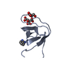
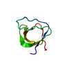
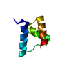
- Structure visualization
| Structure viewer | Molecule: MolmilJmol/JSmol |
|---|
- Downloads & links
-Download
| PDBx/mmCIF format | 1wdx.cif.gz | 59.9 KB | Display | PDBx/mmCIF format |
|---|---|---|---|---|
| PDB format | pdb1wdx.ent.gz | 45.4 KB | Display | PDB format |
| PDBx/mmJSON format | 1wdx.json.gz | Tree view | PDBx/mmJSON format | |
| Others |  Other downloads Other downloads |
-Validation report
| Summary document | 1wdx_validation.pdf.gz | 444.8 KB | Display | wwPDB validaton report |
|---|---|---|---|---|
| Full document | 1wdx_full_validation.pdf.gz | 446.1 KB | Display | |
| Data in XML | 1wdx_validation.xml.gz | 10.3 KB | Display | |
| Data in CIF | 1wdx_validation.cif.gz | 13.7 KB | Display | |
| Arichive directory | https://data.pdbj.org/pub/pdb/validation_reports/wd/1wdxftp://data.pdbj.org/pub/pdb/validation_reports/wd/1wdx | HTTPS FTP |
-Related structure data
| Similar structure data |
|---|
-Links
 PDBj
PDBj- Assembly
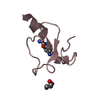
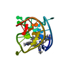
| Deposited unit | 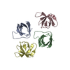
| ||||||||||||||||||||||||||||||
|---|---|---|---|---|---|---|---|---|---|---|---|---|---|---|---|---|---|---|---|---|---|---|---|---|---|---|---|---|---|---|---|
| 1 | 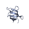
| ||||||||||||||||||||||||||||||
| 2 | 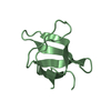
| ||||||||||||||||||||||||||||||
| 3 | 
| ||||||||||||||||||||||||||||||
| 4 | 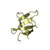
| ||||||||||||||||||||||||||||||
| Unit cell |
| ||||||||||||||||||||||||||||||
| Noncrystallographic symmetry (NCS) | NCS domain:
NCS domain segments: Component-ID: 1 / Ens-ID: 1 / Beg auth comp-ID: PRO / Beg label comp-ID: PRO / End auth comp-ID: VAL / End label comp-ID: VAL / Refine code: 4 / Auth seq-ID: 5 - 67 / Label seq-ID: 5 - 67
|
-Components
| #1: Protein | Mass: 7878.516 Da / Num. of mol.: 4 / Fragment: SH3 domain Source method: isolated from a genetically manipulated source Source: (gene. exp.) Plasmid: pDEST17 / Production host:  #2: Water | ChemComp-HOH / |  Mass: 18.015 Da / Num. of mol.: 16 / Source method: isolated from a natural source / Formula: H2O Mass: 18.015 Da / Num. of mol.: 16 / Source method: isolated from a natural source / Formula: H2O |
|---|
-Experimental details
-Experiment
| Experiment | Method: X-RAY DIFFRACTION / Number of used crystals: 1 |
|---|
- Sample preparation
| Crystal | Density Matthews: 2.1 Å3/Da / Density % sol: 40.6 % |
|---|---|
| Crystal grow | Temperature: 293 K / Method: vapor diffusion, sitting drop Details: 3.2M ammonium sulfate, VAPOR DIFFUSION, SITTING DROP, temperature 293K |
-Data collection
| Diffraction | Mean temperature: 100 K |
|---|---|
| Diffraction source | Source: SYNCHROTRON / Site: EMBL/DESY, HAMBURG  / Beamline: BW7A / Beamline: BW7A |
| Detector | Type: MARRESEARCH / Detector: CCD / Date: Jan 28, 2004 |
| Radiation | Protocol: SINGLE WAVELENGTH / Monochromatic (M) / Laue (L): M / Scattering type: x-ray |
| Radiation wavelength | Relative weight: 1 |
| Reflection | Resolution: 2.2→20 Å / Num. all: 11615 / Num. obs: 11615 / % possible obs: 90.8 % / Observed criterion σ(F): -3 / Observed criterion σ(I): -3 / Redundancy: 1.7 % / Biso Wilson estimate: 18 Å2 / Rsym value: 0.144 / Net I/σ(I): 3.9 |
| Reflection shell | Resolution: 2.2→2.4 Å / Redundancy: 1.7 % / Mean I/σ(I) obs: 2.3 / Num. unique all: 2707 / Rsym value: 0.291 / % possible all: 92.5 |
- Processing
| Software |
| ||||||||||||||||||||||||||||||||||||||||||||||||||||||||||||||||||||||||||||||||||||||||||||||||||||
|---|---|---|---|---|---|---|---|---|---|---|---|---|---|---|---|---|---|---|---|---|---|---|---|---|---|---|---|---|---|---|---|---|---|---|---|---|---|---|---|---|---|---|---|---|---|---|---|---|---|---|---|---|---|---|---|---|---|---|---|---|---|---|---|---|---|---|---|---|---|---|---|---|---|---|---|---|---|---|---|---|---|---|---|---|---|---|---|---|---|---|---|---|---|---|---|---|---|---|---|---|---|
| Refinement | Method to determine structure: MOLECULAR REPLACEMENT Starting model: atomic-resolution structure of yeast bbc1 SH3 domain Resolution: 2.5→10 Å / Cor.coef. Fo:Fc: 0.814 / Cor.coef. Fo:Fc free: 0.729 / SU B: 16.625 / SU ML: 0.377 / Cross valid method: THROUGHOUT / σ(F): -3 / ESU R Free: 0.456 / Stereochemistry target values: Engh & Huber / Details: HYDROGENS HAVE BEEN ADDED IN THE RIDING POSITIONS
| ||||||||||||||||||||||||||||||||||||||||||||||||||||||||||||||||||||||||||||||||||||||||||||||||||||
| Solvent computation | Ion probe radii: 0.8 Å / Shrinkage radii: 0.8 Å / VDW probe radii: 1.4 Å / Solvent model: MASK | ||||||||||||||||||||||||||||||||||||||||||||||||||||||||||||||||||||||||||||||||||||||||||||||||||||
| Displacement parameters | Biso mean: 10.038 Å2
| ||||||||||||||||||||||||||||||||||||||||||||||||||||||||||||||||||||||||||||||||||||||||||||||||||||
| Refinement step | Cycle: LAST / Resolution: 2.5→10 Å
| ||||||||||||||||||||||||||||||||||||||||||||||||||||||||||||||||||||||||||||||||||||||||||||||||||||
| Refine LS restraints |
| ||||||||||||||||||||||||||||||||||||||||||||||||||||||||||||||||||||||||||||||||||||||||||||||||||||
| Refine LS restraints NCS | Ens-ID: 1 / Number: 956 / Refine-ID: X-RAY DIFFRACTION
| ||||||||||||||||||||||||||||||||||||||||||||||||||||||||||||||||||||||||||||||||||||||||||||||||||||
| LS refinement shell | Resolution: 2.5→2.561 Å / Total num. of bins used: 20 /
|
