 Movie
Movie Controller

[English] 日本語
 Yorodumi
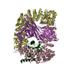
- PDB-1wdl: fatty acid beta-oxidation multienzyme complex from Pseudomonas fr... -
+ Open data

- Basic information
| Entry | Database: PDB / ID: 1wdl | ||||||
|---|---|---|---|---|---|---|---|
| Title | fatty acid beta-oxidation multienzyme complex from Pseudomonas fragi, form II (native4) | ||||||
 Components Components |
| ||||||
 Keywords Keywords | LYASE / OXIDOREDUCTASE/TRANSFERASE / alpha2beta2 heterotetrameric complex / OXIDOREDUCTASE-TRANSFERASE COMPLEX | ||||||
| Function / homology |  Function and homology information Function and homology informationfatty acid beta-oxidation multienzyme complex / 3-hydroxybutyryl-CoA epimerase / 3-hydroxybutyryl-CoA epimerase activity / acetyl-CoA C-acyltransferase / acetyl-CoA C-acyltransferase activity / Delta3-Delta2-enoyl-CoA isomerase / delta(3)-delta(2)-enoyl-CoA isomerase activity / long-chain (3S)-3-hydroxyacyl-CoA dehydrogenase (NAD+) activity / 3-hydroxyacyl-CoA dehydratase activity / 3-hydroxyacyl-CoA dehydrogenase ...fatty acid beta-oxidation multienzyme complex / 3-hydroxybutyryl-CoA epimerase / 3-hydroxybutyryl-CoA epimerase activity / acetyl-CoA C-acyltransferase / acetyl-CoA C-acyltransferase activity / Delta3-Delta2-enoyl-CoA isomerase / delta(3)-delta(2)-enoyl-CoA isomerase activity / long-chain (3S)-3-hydroxyacyl-CoA dehydrogenase (NAD+) activity / 3-hydroxyacyl-CoA dehydratase activity / 3-hydroxyacyl-CoA dehydrogenase / enoyl-CoA hydratase / phenylacetate catabolic process / fatty acid beta-oxidation / NAD+ binding / cytoplasm Similarity search - Function | ||||||
| Biological species |  Pseudomonas fragi (bacteria) Pseudomonas fragi (bacteria) | ||||||
| Method |  X-RAY DIFFRACTION / MOLECULAR REPLACEMENT / Resolution: 3.5 Å X-RAY DIFFRACTION / MOLECULAR REPLACEMENT / Resolution: 3.5 Å | ||||||
 Authors Authors | Ishikawa, M. / Tsuchiya, D. / Oyama, T. / Tsunaka, Y. / Morikawa, K. | ||||||
 Citation Citation | Journal: Embo J. / Year: 2004 Title: Structural basis for channelling mechanism of a fatty acid beta-oxidation multienzyme complex Authors: Ishikawa, M. / Tsuchiya, D. / Oyama, T. / Tsunaka, Y. / Morikawa, K. #1: Journal: Biochem.J. / Year: 1997 Title: Reconstitution, morphology and crystallization of a fatty acid beta-oxidation multienzyme complex from Pseudomonas fragi Authors: Ishikawa, M. / Mikami, Y. / Usukura, J. / Iwasaki, H. / Shinagawa, H. / Morikawa, K. | ||||||
| History |
|
- Structure visualization
| Structure viewer | Molecule: MolmilJmol/JSmol |
|---|
- Downloads & links
-Download
| PDBx/mmCIF format | 1wdl.cif.gz | 374.3 KB | Display | PDBx/mmCIF format |
|---|---|---|---|---|
| PDB format | pdb1wdl.ent.gz | 307 KB | Display | PDB format |
| PDBx/mmJSON format | 1wdl.json.gz | Tree view | PDBx/mmJSON format | |
| Others |  Other downloads Other downloads |
-Validation report
| Arichive directory | https://data.pdbj.org/pub/pdb/validation_reports/wd/1wdlftp://data.pdbj.org/pub/pdb/validation_reports/wd/1wdl | HTTPS FTP |
|---|
-Related structure data


-Links
 PDBj
PDBj


- Assembly
| Deposited unit | 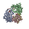
| ||||||||
|---|---|---|---|---|---|---|---|---|---|
| 1 |
| ||||||||
| Unit cell |
| ||||||||
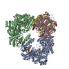
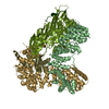
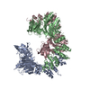
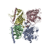
| Details | The biological assembly is a heterotetramer as observed in the asymmetric unit. |
-Components
| #1: Protein | Mass: 77232.266 Da / Num. of mol.: 2 Source method: isolated from a genetically manipulated source Source: (gene. exp.) Pseudomonas fragi (bacteria) / Plasmid: pT7-7 / Production host: References: UniProt: P28793, enoyl-CoA hydratase, Delta3-Delta2-enoyl-CoA isomerase, 3-hydroxyacyl-CoA dehydrogenase, 3-hydroxybutyryl-CoA epimerase #2: Protein | Mass: 41522.730 Da / Num. of mol.: 2 Source method: isolated from a genetically manipulated source Source: (gene. exp.) Pseudomonas fragi (bacteria) / Plasmid: pT7-7 / Production host: #3: Chemical |   Mass: 663.425 Da / Num. of mol.: 2 / Source method: obtained synthetically / Formula: C21H27N7O14P2 / Comment: NAD*YM Mass: 663.425 Da / Num. of mol.: 2 / Source method: obtained synthetically / Formula: C21H27N7O14P2 / Comment: NAD*YM#4: Chemical |   Mass: 350.491 Da / Num. of mol.: 3 / Source method: obtained synthetically / Formula: C18H38O6 / Comment: C8E5, detergent*YM Mass: 350.491 Da / Num. of mol.: 3 / Source method: obtained synthetically / Formula: C18H38O6 / Comment: C8E5, detergent*YM#5: Chemical |   Mass: 809.571 Da / Num. of mol.: 2 / Source method: obtained synthetically / Formula: C23H38N7O17P3S Mass: 809.571 Da / Num. of mol.: 2 / Source method: obtained synthetically / Formula: C23H38N7O17P3S |
|---|
-Experimental details
-Experiment
| Experiment | Method: X-RAY DIFFRACTION / Number of used crystals: 1 |
|---|
- Sample preparation
| Crystal | Density Matthews: 2.29 Å3/Da / Density % sol: 45.8 % |
|---|---|
| Crystal grow | Temperature: 293 K / Method: vapor diffusion, hanging drop / pH: 5.5 Details: PEG4000, MES, n-octylpentaoxyethylene, NAD+, acetoacetyl-CoA, pH 5.5, VAPOR DIFFUSION, HANGING DROP, temperature 293K |
-Data collection
| Diffraction | Mean temperature: 100 K |
|---|---|
| Diffraction source | Source: ROTATING ANODE / Type: MACSCIENCE / Wavelength: 1.5418 Å |
| Detector | Type: MACSCIENCE / Detector: IMAGE PLATE / Date: Oct 5, 1998 |
| Radiation | Monochromator: mirror / Protocol: SINGLE WAVELENGTH / Monochromatic (M) / Laue (L): M / Scattering type: x-ray |
| Radiation wavelength | Wavelength: 1.5418 Å / Relative weight: 1 |
| Reflection | Resolution: 3.5→50 Å / Num. obs: 27768 / % possible obs: 99.5 % / Observed criterion σ(F): 0 / Observed criterion σ(I): 0 / Rmerge(I) obs: 0.157 |
| Reflection shell | Resolution: 3.5→3.72 Å / Rmerge(I) obs: 0.302 / % possible all: 100 |
- Processing
| Software |
| ||||||||||||||||||||
|---|---|---|---|---|---|---|---|---|---|---|---|---|---|---|---|---|---|---|---|---|---|
| Refinement | Method to determine structure: MOLECULAR REPLACEMENT Starting model: form I Resolution: 3.5→12 Å / Cross valid method: THROUGHOUT / σ(F): 2 / Stereochemistry target values: Engh & Huber
| ||||||||||||||||||||
| Refine analyze |
| ||||||||||||||||||||
| Refinement step | Cycle: LAST / Resolution: 3.5→12 Å
| ||||||||||||||||||||
| Refine LS restraints |
| ||||||||||||||||||||
| LS refinement shell | Resolution: 3.5→3.62 Å
|
