 Movie
Movie Controller


+ Open data

- Basic information
| Entry | Database: PDB / ID: 1wbp | ||||||
|---|---|---|---|---|---|---|---|
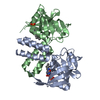
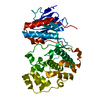
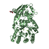
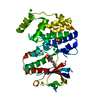
| Title | SRPK1 bound to 9mer docking motif peptide | ||||||
 Components Components |
| ||||||
 Keywords Keywords | TRANSFERASE / SRPK / KINASE / SERINE/THREONINE-PROTEIN KINASE / ATP-BINDING / CHROMOSOME PARTITION / DIFFERENTIATION / MRNA PROCESSING / MRNA SPLICING / NUCLEAR PROTEIN / NUCLEOTIDE-BINDING / PHOSPHORYLATION | ||||||
| Function / homology |  Function and homology information Function and homology informationsperm DNA condensation / positive regulation of cell-cell adhesion / endothelial cell morphogenesis / regulation of mRNA processing / regulation of mRNA splicing, via spliceosome / Maturation of nucleoprotein / negative regulation of viral genome replication / spliceosomal complex assembly / alpha-actinin binding / bicellular tight junction ...sperm DNA condensation / positive regulation of cell-cell adhesion / endothelial cell morphogenesis / regulation of mRNA processing / regulation of mRNA splicing, via spliceosome / Maturation of nucleoprotein / negative regulation of viral genome replication / spliceosomal complex assembly / alpha-actinin binding / bicellular tight junction / positive regulation of viral genome replication / cell projection / Replacement of protamines by nucleosomes in the male pronucleus / RNA splicing / cell periphery / adherens junction / chromosome segregation / nuclear matrix / cell junction / cell-cell junction / protein-containing complex assembly / protein phosphorylation / protein kinase activity / non-specific serine/threonine protein kinase / cell surface receptor signaling pathway / cell adhesion / nuclear speck / intracellular signal transduction / innate immune response / protein serine kinase activity / protein serine/threonine kinase activity / chromatin / magnesium ion binding / signal transduction / RNA binding / nucleoplasm / ATP binding / nucleus / plasma membrane / cytosol / cytoplasm Similarity search - Function | ||||||
| Biological species |  HOMO SAPIENS (human) HOMO SAPIENS (human) | ||||||
| Method |  X-RAY DIFFRACTION / SYNCHROTRON / MOLECULAR REPLACEMENT / Resolution: 2.4 Å X-RAY DIFFRACTION / SYNCHROTRON / MOLECULAR REPLACEMENT / Resolution: 2.4 Å | ||||||
 Authors Authors | Ngo, J.C. / Gullinsgrud, J. / Chakrabarti, S. / Nolen, B. / Aubol, B.E. / Fu, X.-D. / Adams, J.A. / McCammon, J.A. / Ghosh, G. | ||||||
 Citation Citation | Journal: Mol.Cell / Year: 2005 Title: Interplay between Srpk and Clk/Sty Kinases in Phosphorylation of the Splicing Factor Asf/Sf2 is Regulated by a Docking Motif in Asf/Sf2 Authors: Ngo, J.C. / Chakrabarti, S. / Ding, J.-H. / Velazquez-Dones, A. / Nolen, B. / Aubol, B.E. / Adams, J.A. / Fu, X.-D. / Ghosh, G. | ||||||
| History |
|
- Structure visualization
| Structure viewer | Molecule: MolmilJmol/JSmol |
|---|
- Downloads & links
-Download
| PDBx/mmCIF format | 1wbp.cif.gz | 89 KB | Display | PDBx/mmCIF format |
|---|---|---|---|---|
| PDB format | pdb1wbp.ent.gz | 66 KB | Display | PDB format |
| PDBx/mmJSON format | 1wbp.json.gz | Tree view | PDBx/mmJSON format | |
| Others |  Other downloads Other downloads |
-Validation report
| Arichive directory | https://data.pdbj.org/pub/pdb/validation_reports/wb/1wbpftp://data.pdbj.org/pub/pdb/validation_reports/wb/1wbp | HTTPS FTP |
|---|
-Related structure data
| Related structure data |  1wakS S: Starting model for refinement |
|---|---|
| Similar structure data |


-Links
 PDBj
PDBj
- Assembly
| Deposited unit | 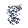
| ||||||||
|---|---|---|---|---|---|---|---|---|---|
| 1 |
| ||||||||
| Unit cell |
| ||||||||
| Details | THE PROTEIN IS A MONOMER IN SOLUTION, BUT SINCEIN THIS ENTRY, IT IS COMPLEX WITH A PEPTIDE, THEENTRY IS BEING MARKED AS DIMERIC.FOR THE HETERO-ASSEMBLY DESCRIBED BY REMARK 350 |
-Components
| #1: Protein | Mass: 45225.598 Da / Num. of mol.: 1 / Fragment: RESIDUES 42-256 AND 474-655 Source method: isolated from a genetically manipulated source Source: (gene. exp.) HOMO SAPIENS (human) / Plasmid: PET15B / Production host:  References: UniProt: Q12890, UniProt: Q96SB4*PLUS, EC: 2.7.1.37 |
|---|---|
| #2: Protein/peptide | Mass: 1218.394 Da / Num. of mol.: 1 / Fragment: RESIDUES 1382-1390 / Source method: obtained synthetically Details: 9 MER PEPTIDE RRRERSPTR BOUND TO A GROOVE FORMED BY THE MAP KINASE INSERT, HELIX AF AND HELIX AG Source: (synth.) HOMO SAPIENS (human) / References: UniProt: Q96QZ7 |
| #3: Chemical | ChemComp-ACT /   Mass: 59.044 Da / Num. of mol.: 1 / Source method: obtained synthetically / Formula: C2H3O2 Mass: 59.044 Da / Num. of mol.: 1 / Source method: obtained synthetically / Formula: C2H3O2 |
| #4: Chemical | ChemComp-ADP /   Mass: 427.201 Da / Num. of mol.: 1 / Source method: obtained synthetically / Formula: C10H15N5O10P2 / Comment: ADP, energy-carrying molecule*YM Mass: 427.201 Da / Num. of mol.: 1 / Source method: obtained synthetically / Formula: C10H15N5O10P2 / Comment: ADP, energy-carrying molecule*YM |
| Compound details | INVOLVED IN THE REGULATORY NETWORK FOR SPLICING, CONTROLLING THE INTRANUCLEAR DISTRIBUTION OF ...INVOLVED IN THE REGULATORY |
| Sequence details | PROTEIN SEQUENCE FROM NCBI (PROTEIN) ACCESSION NP_003128 |
-Experimental details
-Experiment
| Experiment | Method: X-RAY DIFFRACTION / Number of used crystals: 1 |
|---|
- Sample preparation
| Crystal | Density Matthews: 2.99 Å3/Da / Density % sol: 58.82 % | ||||||||||||||||||||||||||||||||||||||||||||||||
|---|---|---|---|---|---|---|---|---|---|---|---|---|---|---|---|---|---|---|---|---|---|---|---|---|---|---|---|---|---|---|---|---|---|---|---|---|---|---|---|---|---|---|---|---|---|---|---|---|---|
| Crystal grow | pH: 7 Details: 100MM SODIUM CITRATE PH5.6, 200MM AMMONIUM ACETATE, 15%PEG3350, 5MM PEPTIDE, 5MM ADP, 10MM MGCL2, pH 7.00 | ||||||||||||||||||||||||||||||||||||||||||||||||
| Crystal grow | *PLUS Temperature: 18 ℃ / pH: 8.5 / Method: vapor diffusion, hanging drop | ||||||||||||||||||||||||||||||||||||||||||||||||
| Components of the solutions | *PLUS
|
-Data collection
| Diffraction | Mean temperature: 105 K |
|---|---|
| Diffraction source | Source: SYNCHROTRON / Site: APS  / Beamline: 19-ID / Wavelength: 1.0332 / Beamline: 19-ID / Wavelength: 1.0332 |
| Detector | Type: ADSC CCD / Detector: CCD / Date: Nov 22, 2003 |
| Radiation | Protocol: SINGLE WAVELENGTH / Monochromatic (M) / Laue (L): M / Scattering type: x-ray |
| Radiation wavelength | Wavelength: 1.0332 Å / Relative weight: 1 |
| Reflection | Resolution: 2.4→30 Å / Num. obs: 22765 / % possible obs: 97.8 % / Observed criterion σ(I): 1 / Redundancy: 11 % / Biso Wilson estimate: 10.9 Å2 / Rmerge(I) obs: 0.07 / Net I/σ(I): 8.1 |
| Reflection shell | Resolution: 2.4→2.49 Å / Redundancy: 7.2 % / Rmerge(I) obs: 0.56 / Mean I/σ(I) obs: 2.9 / % possible all: 84 |
| Reflection | *PLUS Num. measured all: 255743 / Rmerge(I) obs: 0.072 |
| Reflection shell | *PLUS % possible obs: 84 % / Rmerge(I) obs: 0.565 / Mean I/σ(I) obs: 2.86 |
- Processing
| Software |
| ||||||||||||||||||||||||||||||||||||||||||||||||||||||||||||||||||||||||||||||||
|---|---|---|---|---|---|---|---|---|---|---|---|---|---|---|---|---|---|---|---|---|---|---|---|---|---|---|---|---|---|---|---|---|---|---|---|---|---|---|---|---|---|---|---|---|---|---|---|---|---|---|---|---|---|---|---|---|---|---|---|---|---|---|---|---|---|---|---|---|---|---|---|---|---|---|---|---|---|---|---|---|---|
| Refinement | Method to determine structure: MOLECULAR REPLACEMENT Starting model: PDB ENTRY 1WAK Resolution: 2.4→28.26 Å / Rfactor Rfree error: 0.011 / Data cutoff high absF: 384267.63 / Data cutoff low absF: 0 / Isotropic thermal model: RESTRAINED / Cross valid method: THROUGHOUT / σ(F): 2
| ||||||||||||||||||||||||||||||||||||||||||||||||||||||||||||||||||||||||||||||||
| Solvent computation | Solvent model: FLAT MODEL / Bsol: 51.7232 Å2 / ksol: 0.319045 e/Å3 | ||||||||||||||||||||||||||||||||||||||||||||||||||||||||||||||||||||||||||||||||
| Displacement parameters | Biso mean: 96.2 Å2
| ||||||||||||||||||||||||||||||||||||||||||||||||||||||||||||||||||||||||||||||||
| Refine analyze |
| ||||||||||||||||||||||||||||||||||||||||||||||||||||||||||||||||||||||||||||||||
| Refinement step | Cycle: LAST / Resolution: 2.4→28.26 Å
| ||||||||||||||||||||||||||||||||||||||||||||||||||||||||||||||||||||||||||||||||
| Refine LS restraints |
| ||||||||||||||||||||||||||||||||||||||||||||||||||||||||||||||||||||||||||||||||
| LS refinement shell | Resolution: 2.4→2.55 Å / Total num. of bins used: 6
| ||||||||||||||||||||||||||||||||||||||||||||||||||||||||||||||||||||||||||||||||
| Xplor file |
| ||||||||||||||||||||||||||||||||||||||||||||||||||||||||||||||||||||||||||||||||
| Software | *PLUS Name: CNS / Version: 1 / Classification: refinement | ||||||||||||||||||||||||||||||||||||||||||||||||||||||||||||||||||||||||||||||||
| Refinement | *PLUS % reflection Rfree: 5 % | ||||||||||||||||||||||||||||||||||||||||||||||||||||||||||||||||||||||||||||||||
| Solvent computation | *PLUS | ||||||||||||||||||||||||||||||||||||||||||||||||||||||||||||||||||||||||||||||||
| Displacement parameters | *PLUS | ||||||||||||||||||||||||||||||||||||||||||||||||||||||||||||||||||||||||||||||||
| Refine LS restraints | *PLUS
|
