 Movie
Movie Controller

[English] 日本語
 Yorodumi
- PDB-1w74: X-ray structure of peptidyl-prolyl cis-trans isomerase A, PpiA, R... -
+ Open data

- Basic information
| Entry | Database: PDB / ID: 1w74 | ||||||
|---|---|---|---|---|---|---|---|
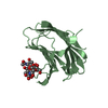
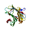
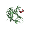
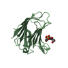
| Title | X-ray structure of peptidyl-prolyl cis-trans isomerase A, PpiA, Rv0009, from Mycobacterium tuberculosis. | ||||||
 Components Components | PEPTIDYL-PROLYL CIS-TRANS ISOMERASE A | ||||||
 Keywords Keywords | ISOMERASE / PEPTIDYL-PROLYL CIS-TRANS ISOMERASE / CYCLOPHILIN / PPIASE / RV0009 / ROTAMASE / STRUCTURAL PROTEOMICS IN EUROPE / SPINE / STRUCTURAL GENOMICS | ||||||
| Function / homology |  Function and homology information Function and homology informationresponse to iron ion / peptidoglycan-based cell wall / peptidylprolyl isomerase / peptidyl-prolyl cis-trans isomerase activity / protein folding / extracellular region / plasma membrane / cytosol / cytoplasm Similarity search - Function | ||||||
| Biological species |   MYCOBACTERIUM TUBERCULOSIS (bacteria) MYCOBACTERIUM TUBERCULOSIS (bacteria) | ||||||
| Method |  X-RAY DIFFRACTION / SYNCHROTRON / MOLECULAR REPLACEMENT / Resolution: 2.6 Å X-RAY DIFFRACTION / SYNCHROTRON / MOLECULAR REPLACEMENT / Resolution: 2.6 Å | ||||||
 Authors Authors | Henriksson, L.M. / Johansson, P. / Unge, T. / Mowbray, S.L. | ||||||
 Citation Citation | Journal: Eur.J.Biochem. / Year: 2004 Title: X-Ray Structure of Peptidyl-Prolyl Cis-Trans Isomerase a from Mycobacterium Tuberculosis Authors: Henriksson, L.M. / Johansson, P. / Unge, T. / Mowbray, S.L. | ||||||
| History |
| ||||||
| Remark 650 | HELIX DETERMINATION METHOD: AUTHOR PROVIDED. | ||||||
| Remark 700 | SHEET DETERMINATION METHOD: DSSP THE SHEETS PRESENTED AS "AA" IN EACH CHAIN ON SHEET RECORDS BELOW ... SHEET DETERMINATION METHOD: DSSP THE SHEETS PRESENTED AS "AA" IN EACH CHAIN ON SHEET RECORDS BELOW IS ACTUALLY AN 9-STRANDED BARREL THIS IS REPRESENTED BY A 10-STRANDED SHEET IN WHICH THE FIRST AND LAST STRANDS ARE IDENTICAL. THE SHEETS PRESENTED AS "BA" IN EACH CHAIN ON SHEET RECORDS BELOW IS ACTUALLY AN 9-STRANDED BARREL THIS IS REPRESENTED BY A 10-STRANDED SHEET IN WHICH THE FIRST AND LAST STRANDS ARE IDENTICAL. |
- Structure visualization
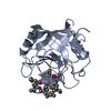
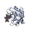
| Structure viewer | Molecule: MolmilJmol/JSmol |
|---|
- Downloads & links
-Download
| PDBx/mmCIF format | 1w74.cif.gz | 74.9 KB | Display | PDBx/mmCIF format |
|---|---|---|---|---|
| PDB format | pdb1w74.ent.gz | 56.6 KB | Display | PDB format |
| PDBx/mmJSON format | 1w74.json.gz | Tree view | PDBx/mmJSON format | |
| Others |  Other downloads Other downloads |
-Validation report
| Arichive directory | https://data.pdbj.org/pub/pdb/validation_reports/w7/1w74ftp://data.pdbj.org/pub/pdb/validation_reports/w7/1w74 | HTTPS FTP |
|---|
-Related structure data
| Related structure data |  1awrS S: Starting model for refinement |
|---|---|
| Similar structure data |

-Links
 PDBj
PDBj

- Assembly
| Deposited unit | 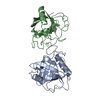
| ||||||||
|---|---|---|---|---|---|---|---|---|---|
| 1 | 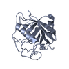
| ||||||||
| 2 | 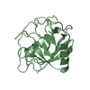
| ||||||||
| Unit cell |
| ||||||||
| Noncrystallographic symmetry (NCS) | NCS oper: (Code: given Matrix: (-0.7467, 0.6651, -0.002), Vector: |
-Components
| #1: Protein | Mass: 20303.445 Da / Num. of mol.: 2 Source method: isolated from a genetically manipulated source Source: (gene. exp.) MYCOBACTERIUM TUBERCULOSIS (bacteria) / Strain: H37RV / Plasmid: PCR T7/CT-TOPO / Production host: References: UniProt: P71578, UniProt: P9WHW3*PLUS, peptidylprolyl isomerase #2: Water | ChemComp-HOH / |  Mass: 18.015 Da / Num. of mol.: 13 / Source method: isolated from a natural source / Formula: H2O Mass: 18.015 Da / Num. of mol.: 13 / Source method: isolated from a natural source / Formula: H2OCompound details | PPIASES ACCELERATE THE FOLDING OF PROTEINS. IT CATALYZES THE CIS-TRANS ISOMERIZATION OF PROLINE ...PPIASES ACCELERATE | Sequence details | THE PROTEIN WAS EXPRESSED WITH AN ADDITIONAL 6-HISTIDINE TAG (MAHHHHHHSG, G REPLACING THE INITIAL M) ...THE PROTEIN WAS EXPRESSED WITH AN ADDITIONAL | |
|---|
-Experimental details
-Experiment
| Experiment | Method: X-RAY DIFFRACTION / Number of used crystals: 1 |
|---|
- Sample preparation
| Crystal | Density Matthews: 3.1 Å3/Da / Density % sol: 60 % |
|---|---|
| Crystal grow | pH: 6 Details: PROTEIN WAS CO-CRYSTALLIZED WITH THE PEPTIDE HAGPIA (FINAL CONCENTRATION 1 MM) FROM 30% PEG-200, 5% PEG-3000, 0.1 M MES-HCL PH 6.0; THEN SOAKED IN THE RESERVOIR SOLUTION PLUS 1 MM HAGPIA. |
-Data collection
| Diffraction | Mean temperature: 110 K |
|---|---|
| Diffraction source | Source: SYNCHROTRON / Site: ESRF  / Beamline: ID14-1 / Wavelength: 0.934 / Beamline: ID14-1 / Wavelength: 0.934 |
| Detector | Type: ADSC CCD / Detector: CCD / Date: Feb 1, 2004 / Details: SAGITALLY FOCUSING GE(220) AND A MULTILAYER |
| Radiation | Monochromator: DIAMOND (111), GE(220) / Protocol: SINGLE WAVELENGTH / Monochromatic (M) / Laue (L): M / Scattering type: x-ray |
| Radiation wavelength | Wavelength: 0.934 Å / Relative weight: 1 |
| Reflection | Resolution: 2.6→32.62 Å / Num. obs: 14896 / % possible obs: 99.6 % / Observed criterion σ(I): 0 / Redundancy: 5.4 % / Rmerge(I) obs: 0.1 / Net I/σ(I): 6.2 |
| Reflection shell | Resolution: 2.6→2.74 Å / Redundancy: 5.4 % / Rmerge(I) obs: 0.59 / Mean I/σ(I) obs: 1.3 / % possible all: 99.8 |
- Processing
| Software |
| ||||||||||||||||||||||||||||||||||||||||||||||||||||||||||||||||||||||||||||||||||||||||||||||||||||||||||||||||||||||||||||||||||||||||||||||||||||||||||||||||||||||||||||||||||||||
|---|---|---|---|---|---|---|---|---|---|---|---|---|---|---|---|---|---|---|---|---|---|---|---|---|---|---|---|---|---|---|---|---|---|---|---|---|---|---|---|---|---|---|---|---|---|---|---|---|---|---|---|---|---|---|---|---|---|---|---|---|---|---|---|---|---|---|---|---|---|---|---|---|---|---|---|---|---|---|---|---|---|---|---|---|---|---|---|---|---|---|---|---|---|---|---|---|---|---|---|---|---|---|---|---|---|---|---|---|---|---|---|---|---|---|---|---|---|---|---|---|---|---|---|---|---|---|---|---|---|---|---|---|---|---|---|---|---|---|---|---|---|---|---|---|---|---|---|---|---|---|---|---|---|---|---|---|---|---|---|---|---|---|---|---|---|---|---|---|---|---|---|---|---|---|---|---|---|---|---|---|---|---|---|
| Refinement | Method to determine structure: MOLECULAR REPLACEMENT Starting model: PDB ENTRY 1AWR Resolution: 2.6→30 Å / Cor.coef. Fo:Fc: 0.945 / Cor.coef. Fo:Fc free: 0.933 / SU B: 9.999 / SU ML: 0.207 / Cross valid method: THROUGHOUT / ESU R: 0.454 / ESU R Free: 0.258 / Stereochemistry target values: MAXIMUM LIKELIHOOD / Details: HYDROGENS HAVE BEEN ADDED IN THE RIDING POSITIONS.
| ||||||||||||||||||||||||||||||||||||||||||||||||||||||||||||||||||||||||||||||||||||||||||||||||||||||||||||||||||||||||||||||||||||||||||||||||||||||||||||||||||||||||||||||||||||||
| Solvent computation | Ion probe radii: 0.8 Å / Shrinkage radii: 0.8 Å / VDW probe radii: 1.4 Å / Solvent model: BABINET MODEL WITH MASK | ||||||||||||||||||||||||||||||||||||||||||||||||||||||||||||||||||||||||||||||||||||||||||||||||||||||||||||||||||||||||||||||||||||||||||||||||||||||||||||||||||||||||||||||||||||||
| Displacement parameters | Biso mean: 61.82 Å2
| ||||||||||||||||||||||||||||||||||||||||||||||||||||||||||||||||||||||||||||||||||||||||||||||||||||||||||||||||||||||||||||||||||||||||||||||||||||||||||||||||||||||||||||||||||||||
| Refinement step | Cycle: LAST / Resolution: 2.6→30 Å
| ||||||||||||||||||||||||||||||||||||||||||||||||||||||||||||||||||||||||||||||||||||||||||||||||||||||||||||||||||||||||||||||||||||||||||||||||||||||||||||||||||||||||||||||||||||||
| Refine LS restraints |
|