 Movie
Movie Controller

[English] 日本語
 Yorodumi
- PDB-1w0c: Inhibition of Leishmania major pteridine reductase (PTR1) by 2,4,... -
+ Open data

- Basic information
| Entry | Database: PDB / ID: 1w0c | ||||||
|---|---|---|---|---|---|---|---|
| Title | Inhibition of Leishmania major pteridine reductase (PTR1) by 2,4,6-triaminoquinazoline; structure of the NADP ternary complex. | ||||||
 Components Components | PTERIDINE REDUCTASE | ||||||
 Keywords Keywords | OXIDOREDUCTASE / ENZYME INHIBITOR / PTERIN / SHORT-CHAIN REDUCTASE / LEISHMANIA / METHOTREXATE / TRYPANOSOMA / NADP / METHOTREXATE RESISTANCE | ||||||
| Function / homology |  Function and homology information Function and homology informationpteridine reductase / 6,7-dihydropteridine reductase activity / pteridine reductase activity / tetrahydrobiopterin biosynthetic process / response to methotrexate / oxidoreductase activity / cytosol Similarity search - Function | ||||||
| Biological species |  LEISHMANIA MAJOR (eukaryote) LEISHMANIA MAJOR (eukaryote) | ||||||
| Method |  X-RAY DIFFRACTION / SYNCHROTRON / MOLECULAR REPLACEMENT / Resolution: 2.6 Å X-RAY DIFFRACTION / SYNCHROTRON / MOLECULAR REPLACEMENT / Resolution: 2.6 Å | ||||||
 Authors Authors | Mcluskey, K. / Gibellini, F. / Carvalho, P. / Avery, M. / Hunter, W. | ||||||
 Citation Citation | Journal: Acta Crystallogr.,Sect.D / Year: 2004 Title: Inhibition of Leishmania Major Pteridine Reductase by 2,4,6-Triaminoquinazoline: Structure of the Nadph Ternary Complex Authors: Mcluskey, K. / Gibellini, F. / Carvalho, P. / Avery, M. / Hunter, W. | ||||||
| History |
|
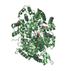
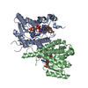
- Structure visualization
| Structure viewer | Molecule: MolmilJmol/JSmol |
|---|
- Downloads & links
-Download
| PDBx/mmCIF format | 1w0c.cif.gz | 432.2 KB | Display | PDBx/mmCIF format |
|---|---|---|---|---|
| PDB format | pdb1w0c.ent.gz | 353.3 KB | Display | PDB format |
| PDBx/mmJSON format | 1w0c.json.gz | Tree view | PDBx/mmJSON format | |
| Others |  Other downloads Other downloads |
-Validation report
| Arichive directory | https://data.pdbj.org/pub/pdb/validation_reports/w0/1w0cftp://data.pdbj.org/pub/pdb/validation_reports/w0/1w0c | HTTPS FTP |
|---|
-Related structure data
| Related structure data | 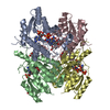 1e92S S: Starting model for refinement |
|---|---|
| Similar structure data |


-Links
 PDBj
PDBj


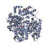
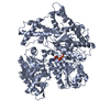
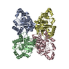
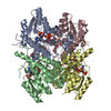
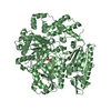
- Assembly
| Deposited unit | 
| ||||||||
|---|---|---|---|---|---|---|---|---|---|
| 1 | 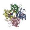
| ||||||||
| 2 | 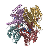
| ||||||||
| Unit cell |
| ||||||||
| Noncrystallographic symmetry (NCS) | NCS oper: (Code: given Matrix: (-1, 0.00046, 0.00029), Vector: |
-Components
| #1: Protein | Mass: 32646.971 Da / Num. of mol.: 8 Source method: isolated from a genetically manipulated source Details: CONTAINS INHIBITOR TAQ / Source: (gene. exp.) LEISHMANIA MAJOR (eukaryote) / Strain: B834 / Production host:  #2: Chemical | ChemComp-NAP /   Mass: 743.405 Da / Num. of mol.: 8 / Source method: obtained synthetically / Formula: C21H28N7O17P3 Mass: 743.405 Da / Num. of mol.: 8 / Source method: obtained synthetically / Formula: C21H28N7O17P3#3: Chemical | ChemComp-TAQ /   Mass: 175.191 Da / Num. of mol.: 9 / Source method: obtained synthetically / Formula: C8H9N5 Mass: 175.191 Da / Num. of mol.: 9 / Source method: obtained synthetically / Formula: C8H9N5#4: Water | ChemComp-HOH / |  Mass: 18.015 Da / Num. of mol.: 1117 / Source method: isolated from a natural source / Formula: H2O Mass: 18.015 Da / Num. of mol.: 1117 / Source method: isolated from a natural source / Formula: H2OCompound details | 5,6,7,8-TETRAHYDRO | |
|---|
-Experimental details
-Experiment
| Experiment | Method: X-RAY DIFFRACTION / Number of used crystals: 1 |
|---|
- Sample preparation
| Crystal | Density Matthews: 2.8 Å3/Da / Density % sol: 56 % |
|---|---|
| Crystal grow | pH: 5.4 Details: 11-14% PEG 5000, 100 MM NAAC PH 5.5 AND 40-140 MM CAAC. |
-Data collection
| Diffraction | Mean temperature: 100 K |
|---|---|
| Diffraction source | Source: SYNCHROTRON / Site: ESRF  / Beamline: ID14-4 / Wavelength: 0.9393 / Beamline: ID14-4 / Wavelength: 0.9393 |
| Detector | Type: ADSC CCD / Detector: CCD / Date: Sep 15, 2003 / Details: TOROIDAL MIRROR |
| Radiation | Monochromator: DOUBLE CRYSTAL, SI(111) OR SI(311) / Protocol: SINGLE WAVELENGTH / Monochromatic (M) / Laue (L): M / Scattering type: x-ray |
| Radiation wavelength | Wavelength: 0.9393 Å / Relative weight: 1 |
| Reflection | Resolution: 1.75→30 Å / Num. obs: 59475 / % possible obs: 91 % / Redundancy: 3.9 % / Biso Wilson estimate: 33 Å2 / Rmerge(I) obs: 0.146 / Net I/σ(I): 6.9 |
| Reflection shell | Resolution: 2.6→2.7 Å / Rmerge(I) obs: 0.406 / Mean I/σ(I) obs: 2.8 / % possible all: 85.2 |
- Processing
| Software |
| ||||||||||||||||||||
|---|---|---|---|---|---|---|---|---|---|---|---|---|---|---|---|---|---|---|---|---|---|
| Refinement | Method to determine structure: MOLECULAR REPLACEMENT Starting model: PDB ENTRY 1E92 Resolution: 2.6→30 Å / SU B: 15.271 / SU ML: 0.331 / Cross valid method: THROUGHOUT / σ(F): 0 / ESU R: 2.3 / ESU R Free: 0.455 Details: THE C-CENTRED ORTHORHOMBIC SPACE GROUP C222 WAS ALSO CONSIDERED BUT THE DATA DID NOT SCALE IN THIS SPACE GROUP.
| ||||||||||||||||||||
| Displacement parameters | Biso mean: 20 Å2
| ||||||||||||||||||||
| Refinement step | Cycle: LAST / Resolution: 2.6→30 Å
|
