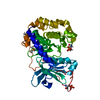
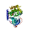
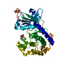
Entry Database : PDB / ID : 1vzoTitle The structure of the N-terminal kinase domain of MSK1 reveals a novel autoinhibitory conformation for a dual kinase protein RIBOSOMAL PROTEIN S6 KINASE ALPHA 5 Keywords / / / Function / homology Function Domain/homology Component
/ / / / / / / / / / / / / / / / / / / / / / / / / / / / / / / / / / / / / / / / / / / / / / / / / / / / / / / / / / / / / / / / / / / / / / Biological species HOMO SAPIENS (human)Method / / / Resolution : 1.8 Å Authors Smith, K.J. / Carter, P.S. / Bridges, A. / Horrocks, P. / Lewis, C. / Pettman, G. / Clarke, A. / Brown, M. / Hughes, J. / Wilkinson, M. ...Smith, K.J. / Carter, P.S. / Bridges, A. / Horrocks, P. / Lewis, C. / Pettman, G. / Clarke, A. / Brown, M. / Hughes, J. / Wilkinson, M. / Bax, B. / Reith, A. Journal : Structure / Year : 2004Title : The Structure of Msk1 Reveals a Novel Autoinhibitory Conformation for a Dual Kinase ProteinAuthors : Smith, K.J. / Carter, P.S. / Bridges, A. / Horrocks, P. / Lewis, C. / Pettman, G. / Clarke, A. / Brown, M. / Hughes, J. / Wilkinson, M. / Bax, B. / Reith, A. History Deposition May 21, 2004 Deposition site / Processing site Revision 1.0 Jun 11, 2004 Provider / Type Revision 1.1 May 8, 2011 Group Revision 1.2 Jul 13, 2011 Group Revision 1.3 Dec 13, 2023 Group Data collection / Database references ... Data collection / Database references / Other / Refinement description Category chem_comp_atom / chem_comp_bond ... chem_comp_atom / chem_comp_bond / database_2 / pdbx_database_status / pdbx_initial_refinement_model Item / _database_2.pdbx_database_accession / _pdbx_database_status.status_code_sf
Show all Show less
 Movie
Movie Controller

 Yorodumi
 Open data
 Basic information
 Components
 Keywords
 Function and homology information
 HOMO SAPIENS (human)
 X-RAY DIFFRACTION /
  Authors
 Citation
 Structure visualization
 Downloads & links
 Other downloads
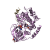


 PDBj
PDBj


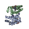
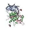
 Assembly
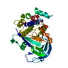

 SPODOPTERA FRUGIPERDA (fall armyworm) / References: UniProt: O75582, EC: 2.7.1.37
 Mass: 78.133 Da / Num. of mol.: 1 / Source method: obtained synthetically / Formula: C2H6OS
 Mass: 96.063 Da / Num. of mol.: 1 / Source method: obtained synthetically / Formula: SO4
 Mass: 18.015 Da / Num. of mol.: 182 / Source method: isolated from a natural source / Formula: H2O
 Sample preparation
 / Beamline: ID14-2 / Wavelength: 0.9326
  Processing