 Movie
Movie Controller

[English] 日本語
 Yorodumi
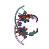
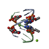
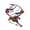
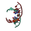
- PDB-1vtb: THE HEXAGONAL CRYSTAL STRUCTURE OF THE A-DNA OCTAMER D(GTGTACAC) ... -
+ Open data

- Basic information
| Entry | Database: PDB / ID: 1vtb | ||||||||||||||||||
|---|---|---|---|---|---|---|---|---|---|---|---|---|---|---|---|---|---|---|---|
| Title | THE HEXAGONAL CRYSTAL STRUCTURE OF THE A-DNA OCTAMER D(GTGTACAC) AND ITS COMPARISON WITH THE TETRAGONAL STRUCTURE CORRELATED VARIATIONS IN HELICAL PARAMETERS | ||||||||||||||||||
 Components Components | DNA (5'-D(* Keywords KeywordsDNA / A-DNA / DOUBLE HELIX | Function / homology | DNA |  Function and homology information Function and homology informationMethod |  X-RAY DIFFRACTION / Resolution: 2 Å X-RAY DIFFRACTION / Resolution: 2 Å  Authors AuthorsJain, S.C. / Zon, G. / Sundaralingam, M. |  CitationJournal: Biochemistry / Year: 1991 CitationJournal: Biochemistry / Year: 1991Title: The Hexagonal Crystal Structure of the A-DNA Octamer d(GTGTACAC) and Its Comparison with the Tetragonal Structure Correlated Variations in Helical Parameters Authors: Jain, S.C. / Zon, G. / Sundaralingam, M. #1: Journal: J.Biol.Chem. / Year: 1989Title: Effect of Crystal Packing Environment on Conformation of the DNA Duplex Authors: Jain, S. / Sundaralingam, M. #2: Journal: Biochemistry / Year: 1989Title: Base Only Binding of Spermine in the Deep Groove of the A-DNA Octamer d(GTGTACAC) Authors: Jain, S. / Zon, G. / Sundaralingam, M. History |
|
- Structure visualization
| Structure viewer | Molecule: MolmilJmol/JSmol |
|---|
- Downloads & links
-Download
| PDBx/mmCIF format | 1vtb.cif.gz | 14.1 KB | Display | PDBx/mmCIF format |
|---|---|---|---|---|
| PDB format | pdb1vtb.ent.gz | 8.3 KB | Display | PDB format |
| PDBx/mmJSON format | 1vtb.json.gz | Tree view | PDBx/mmJSON format | |
| Others |  Other downloads Other downloads |
-Validation report
| Summary document | 1vtb_validation.pdf.gz | 312.7 KB | Display | wwPDB validaton report |
|---|---|---|---|---|
| Full document | 1vtb_full_validation.pdf.gz | 315.1 KB | Display | |
| Data in XML | 1vtb_validation.xml.gz | 1.6 KB | Display | |
| Data in CIF | 1vtb_validation.cif.gz | 2.2 KB | Display | |
| Arichive directory | https://data.pdbj.org/pub/pdb/validation_reports/vt/1vtbftp://data.pdbj.org/pub/pdb/validation_reports/vt/1vtb | HTTPS FTP |
-Related structure data
| Similar structure data |
|---|


-Links
 PDBj
PDBj

- Assembly
| Deposited unit | 
| ||||||||
|---|---|---|---|---|---|---|---|---|---|
| 1 | 
| ||||||||
| Unit cell |
|
-Components
| #1: DNA chain | Mass: 2426.617 Da / Num. of mol.: 1 / Source method: obtained synthetically |
|---|---|
| #2: Water | ChemComp-HOH /  Mass: 18.015 Da / Num. of mol.: 50 / Source method: isolated from a natural source / Formula: H2O Mass: 18.015 Da / Num. of mol.: 50 / Source method: isolated from a natural source / Formula: H2O |
-Experimental details
-Experiment
| Experiment | Method: X-RAY DIFFRACTION |
|---|
- Sample preparation
| Crystal | Density Matthews: 2.47 Å3/Da / Density % sol: 50.29 % | |||||||||||||||||||||||||||||||||||||||||||||||||
|---|---|---|---|---|---|---|---|---|---|---|---|---|---|---|---|---|---|---|---|---|---|---|---|---|---|---|---|---|---|---|---|---|---|---|---|---|---|---|---|---|---|---|---|---|---|---|---|---|---|---|
| Crystal grow | Temperature: 278 K / Method: vapor diffusion / Details: VAPOR DIFFUSION, temperature 278.00K | |||||||||||||||||||||||||||||||||||||||||||||||||
| Components of the solutions |
|
-Data collection
| Diffraction source | Source: ROTATING ANODE |
|---|---|
| Detector | Type: ARNDT-WONACOTT / Detector: OSCILLATION CAMERA |
| Radiation | Protocol: SINGLE WAVELENGTH / Monochromatic (M) / Laue (L): M / Scattering type: x-ray |
| Radiation wavelength | Relative weight: 1 |
| Reflection | Highest resolution: 2 Å / Num. all: 8156 / Num. obs: 1683 / Observed criterion σ(I): 1.5 |
- Processing
| Software | Name: NUCLSQ / Classification: refinement | ||||||||||||
|---|---|---|---|---|---|---|---|---|---|---|---|---|---|
| Refinement | Highest resolution: 2 Å / σ(I): 1.5 /
| ||||||||||||
| Refinement step | Cycle: LAST / Highest resolution: 2 Å
|
