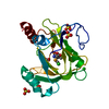
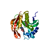
Mass: 23485.158 Da / Num. of mol.: 1 Source method: isolated from a genetically manipulated source Source: (gene. exp.) Mus musculus (house mouse) / Production host: Escherichia coli (E. coli) References: UniProt: Q99JT9, Oxidoreductases; Acting on single donors with incorporation of molecular oxygen (oxygenases); With incorporation of two atoms of oxygen
Resolution: 2.06→28.54 Å / Num. obs: 23148 / % possible obs: 100 % / Redundancy: 6.8 % / Biso Wilson estimate: 43.37 Å2 / Rsym value: 0.101 / Net I/σ(I): 13.6
Reflection shell
Resolution: 2.06→2.17 Å / Redundancy: 5.4 % / Mean I/σ(I) obs: 2.2 / Num. unique all: 3314 / Rsym value: 0.864 / % possible all: 100
-
Processing
Software
Name
Version
Classification
XDS
datascaling
SCALA
5.0)
datascaling
autoSHARP
phasing
REFMAC
5.2.0005
refinement
XDS
datareduction
CCP4
(SCALA)
datascaling
Refinement
Method to determine structure: MAD / Resolution: 2.06→28.53 Å / Cor.coef. Fo:Fc: 0.963 / Cor.coef. Fo:Fc free: 0.949 / SU B: 5.439 / SU ML: 0.081 / TLS residual ADP flag: LIKELY RESIDUAL / Cross valid method: THROUGHOUT / ESU R: 0.115 / ESU R Free: 0.115 Stereochemistry target values: MAXIMUM LIKELIHOOD WITH PHASES Details: 1. HYDROGENS HAVE BEEN ADDED IN THE RIDING POSITIONS 2. THE IDENTITY OF THE METAL BOUND IS UNKNOWN. IT WAS TENTATIVELY ASSIGNED AS A NICKLE. BASED ON INFORMATION OF HOMOLOGOUS PROTEINS, IT ...Details: 1. HYDROGENS HAVE BEEN ADDED IN THE RIDING POSITIONS 2. THE IDENTITY OF THE METAL BOUND IS UNKNOWN. IT WAS TENTATIVELY ASSIGNED AS A NICKLE. BASED ON INFORMATION OF HOMOLOGOUS PROTEINS, IT COULD BE NI2+ OR FE2+. HOWEVER, IT COULD BE ALSO OTHER METALS. 3. SUSPICIOUS DENSITIES: NEAR THR11, IT WAS MODELLED AS A WATER WAT136; ANOTHER DENSITY IN THE ACTIVE SITE WAS MODELLED AS UNKNOWN LIGAND, UNL. THE UNL INTERACTS WITH THE UNKNOWN METAL.
Rfactor
Num. reflection
% reflection
Selection details
Rfree
0.19532
1183
5.1 %
RANDOM
Rwork
0.16229
-
-
-
obs
0.16387
21908
99.94 %
-
Solvent computation
Ion probe radii: 0.8 Å / Shrinkage radii: 0.8 Å / VDW probe radii: 1.2 Å / Solvent model: BABINET MODEL WITH MASK
Displacement parameters
Biso mean: 37.722 Å2
Baniso -1
Baniso -2
Baniso -3
1-
-0.48 Å2
0 Å2
0 Å2
2-
-
-0.48 Å2
0 Å2
3-
-
-
0.96 Å2
Refinement step
Cycle: LAST / Resolution: 2.06→28.53 Å
Protein
Nucleic acid
Ligand
Solvent
Total
Num. atoms
1484
0
14
169
1667
Refine LS restraints
Refine-ID
Type
Dev ideal
Dev ideal target
Number
X-RAY DIFFRACTION
r_bond_refined_d
0.018
0.022
1583
X-RAY DIFFRACTION
r_bond_other_d
0.002
0.02
1421
X-RAY DIFFRACTION
r_angle_refined_deg
1.436
1.949
2143
X-RAY DIFFRACTION
r_angle_other_deg
0.799
3
3295
X-RAY DIFFRACTION
r_dihedral_angle_1_deg
6.425
5
178
X-RAY DIFFRACTION
r_dihedral_angle_2_deg
35.216
23.182
88
X-RAY DIFFRACTION
r_dihedral_angle_3_deg
13.935
15
284
X-RAY DIFFRACTION
r_dihedral_angle_4_deg
11.433
15
16
X-RAY DIFFRACTION
r_chiral_restr
0.083
0.2
221
X-RAY DIFFRACTION
r_gen_planes_refined
0.006
0.02
1729
X-RAY DIFFRACTION
r_gen_planes_other
0.001
0.02
346
X-RAY DIFFRACTION
r_nbd_refined
0.214
0.2
293
X-RAY DIFFRACTION
r_nbd_other
0.196
0.2
1423
X-RAY DIFFRACTION
r_nbtor_other
0.085
0.2
919
X-RAY DIFFRACTION
r_xyhbond_nbd_refined
0.202
0.2
140
X-RAY DIFFRACTION
r_symmetry_vdw_refined
0.107
0.2
6
X-RAY DIFFRACTION
r_symmetry_vdw_other
0.25
0.2
24
X-RAY DIFFRACTION
r_symmetry_hbond_refined
0.153
0.2
5
X-RAY DIFFRACTION
r_mcbond_it
2.887
3
940
X-RAY DIFFRACTION
r_mcbond_other
0.6
3
357
X-RAY DIFFRACTION
r_mcangle_it
3.504
5
1479
X-RAY DIFFRACTION
r_scbond_it
6.033
8
761
X-RAY DIFFRACTION
r_scangle_it
8.382
11
664
X-RAY DIFFRACTION
r_nbtor_refined
0.188
0.2
758
LS refinement shell
Resolution: 2.06→2.114 Å / Total num. of bins used: 20
Rfactor
Num. reflection
% reflection
Rfree
0.291
91
5.43 %
Rwork
0.243
1585
-
obs
-
-
100 %
Refinement TLS params.
Method: refined / Origin x: 45.816 Å / Origin y: 14.8274 Å / Origin z: 49.5691 Å
11
12
13
21
22
23
31
32
33
T
-0.1005 Å2
0.0133 Å2
0.0167 Å2
-
-0.0814 Å2
-0.0269 Å2
-
-
-0.0741 Å2
L
2.4355 °2
-0.1448 °2
-1.1836 °2
-
0.8176 °2
0.2589 °2
-
-
1.8039 °2
S
0.003 Å °
-0.0569 Å °
-0.0636 Å °
0.0065 Å °
0.0336 Å °
-0.0037 Å °
-0.0112 Å °
0.1229 Å °
-0.0366 Å °
Refinement TLS group
Selection: ALL
+
About Yorodumi
-
News
-
Feb 9, 2022. New format data for meta-information of EMDB entries
New format data for meta-information of EMDB entries
Version 3 of the EMDB header file is now the official format.
The previous official version 1.9 will be removed from the archive.
In the structure databanks used in Yorodumi, some data are registered as the other names, "COVID-19 virus" and "2019-nCoV". Here are the details of the virus and the list of structure data.
Jan 31, 2019. EMDB accession codes are about to change! (news from PDBe EMDB page)
EMDB accession codes are about to change! (news from PDBe EMDB page)
The allocation of 4 digits for EMDB accession codes will soon come to an end. Whilst these codes will remain in use, new EMDB accession codes will include an additional digit and will expand incrementally as the available range of codes is exhausted. The current 4-digit format prefixed with “EMD-” (i.e. EMD-XXXX) will advance to a 5-digit format (i.e. EMD-XXXXX), and so on. It is currently estimated that the 4-digit codes will be depleted around Spring 2019, at which point the 5-digit format will come into force.
The EM Navigator/Yorodumi systems omit the EMD- prefix.
Related info.:Q: What is EMD? / ID/Accession-code notation in Yorodumi/EM Navigator
Yorodumi is a browser for structure data from EMDB, PDB, SASBDB, etc.
This page is also the successor to EM Navigator detail page, and also detail information page/front-end page for Omokage search.
The word "yorodu" (or yorozu) is an old Japanese word meaning "ten thousand". "mi" (miru) is to see.
Related info.:EMDB / PDB / SASBDB / Comparison of 3 databanks / Yorodumi Search / Aug 31, 2016. New EM Navigator & Yorodumi / Yorodumi Papers / Jmol/JSmol / Function and homology information / Changes in new EM Navigator and Yorodumi
 Movie
Movie Controller

 Yorodumi
 Open data
 Basic information
 Components
 Keywords
 Function and homology information
 X-RAY DIFFRACTION /
  Authors
 Citation
 Structure visualization
 Downloads & links
 Other downloads
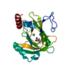


 PDBj
PDBj


 Assembly
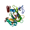


 Mass: 58.693 Da / Num. of mol.: 1 / Source method: obtained synthetically / Formula: Ni
 Mass: 60.095 Da / Num. of mol.: 1 / Source method: obtained synthetically / Formula: C3H8O
 Mass: 18.015 Da / Num. of mol.: 169 / Source method: isolated from a natural source / Formula: H2O
 Sample preparation
 / Beamline: 5.0.2 / Wavelength: 0.979735,0.979562,0.956885
 Processing