 Movie
Movie Controller


+ Open data

- Basic information
| Entry | Database: PDB / ID: 1vpn | ||||||
|---|---|---|---|---|---|---|---|
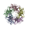
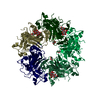
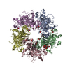
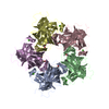
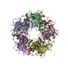
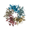
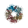
| Title | UNASSEMBLED POLYOMAVIRUS VP1 PENTAMER | ||||||
 Components Components | POLYOMAVIRUS VP1 PENTAMER | ||||||
 Keywords Keywords | VIRAL PROTEIN / VIRUS COAT PROTEIN / VIRUS ASSEMBLY | ||||||
| Function / homology |  Function and homology information Function and homology informationcaveolin-mediated endocytosis of virus by host cell / T=7 icosahedral viral capsid / virion attachment to host cell / host cell nucleus / structural molecule activity Similarity search - Function | ||||||
| Biological species |  Murine polyomavirus Murine polyomavirus | ||||||
| Method |  X-RAY DIFFRACTION / SYNCHROTRON / MOLECULAR REPLACEMENT / Resolution: 2 Å X-RAY DIFFRACTION / SYNCHROTRON / MOLECULAR REPLACEMENT / Resolution: 2 Å | ||||||
 Authors Authors | Stehle, T. / Harrison, S.C. | ||||||
 Citation Citation | Journal: Embo J. / Year: 1997 Title: High-resolution structure of a polyomavirus VP1-oligosaccharide complex: implications for assembly and receptor binding. Authors: Stehle, T. / Harrison, S.C. #1: Journal: Structure / Year: 1996Title: The Structure of Simian Virus 40 Refined at 3.1 A Resolution Authors: Stehle, T. / Gamblin, S.J. / Yan, Y. / Harrison, S.C. #2: Journal: Structure / Year: 1996Title: Crystal Structures of Murine Polyomavirus in Complex with Straight-Chain and Branched-Chain Sialyloligosaccharide Receptor Fragments Authors: Stehle, T. / Harrison, S.C. #3: Journal: Nature / Year: 1994Title: Structure of Murine Polyomavirus Complexed with an Oligosaccharide Receptor Fragment Authors: Stehle, T. / Yan, Y. / Benjamin, T.L. / Harrison, S.C. | ||||||
| History |
|
- Structure visualization
| Structure viewer | Molecule: MolmilJmol/JSmol |
|---|
- Downloads & links
-Download
| PDBx/mmCIF format | 1vpn.cif.gz | 328.9 KB | Display | PDBx/mmCIF format |
|---|---|---|---|---|
| PDB format | pdb1vpn.ent.gz | 266.2 KB | Display | PDB format |
| PDBx/mmJSON format | 1vpn.json.gz | Tree view | PDBx/mmJSON format | |
| Others |  Other downloads Other downloads |
-Validation report
| Arichive directory | https://data.pdbj.org/pub/pdb/validation_reports/vp/1vpnftp://data.pdbj.org/pub/pdb/validation_reports/vp/1vpn | HTTPS FTP |
|---|
-Related structure data
| Related structure data | 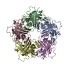 1vpsC  1sidS S: Starting model for refinement C: citing same article ( |
|---|---|
| Similar structure data |


-Links
 PDBj
PDBj


- Assembly
| Deposited unit | 
| ||||||||
|---|---|---|---|---|---|---|---|---|---|
| 1 |
| ||||||||
| 2 | 
| ||||||||
| Unit cell |
| ||||||||
| Details | THE COORDINATE SET CONSISTS OF FIVE VP1 MOLECULES THAT FORM A PENTAMER. |
-Components
| #1: Protein | Mass: 32087.213 Da / Num. of mol.: 5 Source method: isolated from a genetically manipulated source Source: (gene. exp.) Murine polyomavirus / Genus: Polyomavirus / Production host:  #2: Water | ChemComp-HOH / |  Mass: 18.015 Da / Num. of mol.: 2096 / Source method: isolated from a natural source / Formula: H2O Mass: 18.015 Da / Num. of mol.: 2096 / Source method: isolated from a natural source / Formula: H2O |
|---|
-Experimental details
-Experiment
| Experiment | Method: X-RAY DIFFRACTION / Number of used crystals: 1 |
|---|
- Sample preparation
| Crystal | Density Matthews: 4.4 Å3/Da / Density % sol: 65 % | ||||||||||||||||||||||||||||||
|---|---|---|---|---|---|---|---|---|---|---|---|---|---|---|---|---|---|---|---|---|---|---|---|---|---|---|---|---|---|---|---|
| Crystal grow | pH: 8 Details: DROP: 1.0 M AMMONIUM PHOSPHATE PH 8.0 2.5 & ETHANOL 8-10 MG/ML PROTEIN RESERVOIR: 2.0 M AMMONIUM PHOSPHATE PH 8.0 5 % ETHANOL | ||||||||||||||||||||||||||||||
| Crystal | *PLUS Density % sol: 65 % | ||||||||||||||||||||||||||||||
| Crystal grow | *PLUS Method: vapor diffusion, hanging drop | ||||||||||||||||||||||||||||||
| Components of the solutions | *PLUS
|
-Data collection
| Diffraction | Mean temperature: 120 K |
|---|---|
| Diffraction source | Source: SYNCHROTRON / Site: CHESS  / Beamline: F1 / Wavelength: 0.918 / Beamline: F1 / Wavelength: 0.918 |
| Detector | Type: FUJI / Detector: IMAGE PLATE / Date: Dec 1, 1994 / Details: MIRROR |
| Radiation | Monochromator: WIGGLER / Monochromatic (M) / Laue (L): M / Scattering type: x-ray |
| Radiation wavelength | Wavelength: 0.918 Å / Relative weight: 1 |
| Reflection | Resolution: 2→25 Å / Num. obs: 172107 / % possible obs: 83.2 % / Observed criterion σ(I): 0 / Rsym value: 0.106 / Net I/σ(I): 14.9 |
| Reflection shell | Resolution: 2→2.1 Å / Mean I/σ(I) obs: 2.1 / Rsym value: 0.302 / % possible all: 62.2 |
| Reflection | *PLUS Rmerge(I) obs: 0.106 |
- Processing
| Software |
| ||||||||||||||||||||||||||||||||||||||||||||||||||||||||||||
|---|---|---|---|---|---|---|---|---|---|---|---|---|---|---|---|---|---|---|---|---|---|---|---|---|---|---|---|---|---|---|---|---|---|---|---|---|---|---|---|---|---|---|---|---|---|---|---|---|---|---|---|---|---|---|---|---|---|---|---|---|---|
| Refinement | Method to determine structure: MOLECULAR REPLACEMENT Starting model: POLYOMAVIRUS VP1 PENTAMER (PDB ENTRY 1SID) Resolution: 2→20 Å / Isotropic thermal model: RESTRAINED / Cross valid method: THROUGHOUT / σ(F): 0
| ||||||||||||||||||||||||||||||||||||||||||||||||||||||||||||
| Displacement parameters | Biso mean: 29.9 Å2 | ||||||||||||||||||||||||||||||||||||||||||||||||||||||||||||
| Refine analyze | Luzzati coordinate error obs: 0.15 Å / Luzzati d res low obs: 20 Å | ||||||||||||||||||||||||||||||||||||||||||||||||||||||||||||
| Refinement step | Cycle: LAST / Resolution: 2→20 Å
| ||||||||||||||||||||||||||||||||||||||||||||||||||||||||||||
| Refine LS restraints |
| ||||||||||||||||||||||||||||||||||||||||||||||||||||||||||||
| Refine LS restraints NCS | NCS model details: UNRESTRAINED | ||||||||||||||||||||||||||||||||||||||||||||||||||||||||||||
| Xplor file |
|
