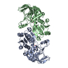
Component-ID: 1 / Ens-ID: 1 / Beg auth comp-ID: MET / Beg label comp-ID: MET / End auth comp-ID: ARG / End label comp-ID: ARG / Refine code: 2 / Auth seq-ID: 1 - 321 / Label seq-ID: 13 - 333
Dom-ID
Auth asym-ID
Label asym-ID
1
A
A
2
B
B
-
Components
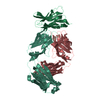
#1: Protein
alaninedehydrogenase
Mass: 36331.465 Da / Num. of mol.: 2 Source method: isolated from a genetically manipulated source Source: (gene. exp.) Archaeoglobus fulgidus (archaea) / Strain: DSM 4304 / Gene: AF1665, arcB / Production host: Escherichia coli (E. coli) / References: UniProt: O28608, alanine dehydrogenase
-
Experimental details
-
Experiment
Experiment
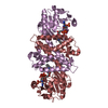
Method: X-RAY DIFFRACTION / Number of used crystals: 2
-
Sample preparation
Crystal
ID
Density Matthews (Å3/Da)
Density % sol (%)
Description
1
2.38
48.22
2
2.7
54.03
DATA FROM A CRYSTAL CONTAINING SE-MET SUBSTITUTED WAS USED FOR THE MAD PHASING EXPERIMENTS AND DATA FROM A CRYSTAL OF THE NATIVE PROTEIN WAS USED FOR THE FINAL REFINEMENT. THE 3 ANGSTROM MAD PHASES WERE EXTENDED TO 2.8 ANGSTROMS USING THE NATIVE STRUCTURE FACTOR AMPLITUDES IN THE PROGRAM RESOLVE. AFTER THE INITIAL TRACE PDB ENTRY 1OMO WAS USED TO FACILITATE COMPLETION OF THE MODEL.
Resolution: 2.8→56.7 Å / Num. obs: 17713 / % possible obs: 99.9 % / Redundancy: 3.7 % / Biso Wilson estimate: 84.89 Å2 / Rmerge(I) obs: 0.057 / Net I/σ(I): 14.8
Reflection shell
Resolution: 2.8→2.87 Å / Redundancy: 3.3 % / Rmerge(I) obs: 0.362 / Mean I/σ(I) obs: 2.3 / Num. unique all: 1280 / % possible all: 99.8
-
Processing
Software
Name
Version
Classification
MOSFLM
datareduction
SCALA
4.2)
datascaling
SOLVE
phasing
RESOLVE
modelbuilding
REFMAC
5.2.0005
refinement
CCP4
(SCALA)
datascaling
RESOLVE
phasing
Refinement
Method to determine structure: MAD / Resolution: 2.8→52.09 Å / Cor.coef. Fo:Fc: 0.943 / Cor.coef. Fo:Fc free: 0.929 / SU B: 37.347 / SU ML: 0.317 / TLS residual ADP flag: LIKELY RESIDUAL / Cross valid method: THROUGHOUT / ESU R Free: 0.378 Stereochemistry target values: MAXIMUM LIKELIHOOD WITH PHASES Details: THE DATA USED IN THE FINAL REFINEMENT WAS FROM A NATIVE CRYSTAL. THE REFINEMENT OF THE COORDINATES WAS RESTRAINED WITH THE EXPERIMENTAL PHASES FROM A CRYSTAL OF THE SELENOMETHIONINE- ...Details: THE DATA USED IN THE FINAL REFINEMENT WAS FROM A NATIVE CRYSTAL. THE REFINEMENT OF THE COORDINATES WAS RESTRAINED WITH THE EXPERIMENTAL PHASES FROM A CRYSTAL OF THE SELENOMETHIONINE-SUBSTITUTED PROTEIN THAT WAS USED FOR INITIAL PHASE DETERMINATION BY MULTIPLE WAVELENGTH ANOMALOUS DISPERSION.
Rfactor
Num. reflection
% reflection
Selection details
Rfree
0.2539
897
5.1 %
RANDOM
Rwork
0.21742
-
-
-
obs
0.21926
16757
99.79 %
-
Solvent computation
Ion probe radii: 0.8 Å / Shrinkage radii: 0.8 Å / VDW probe radii: 1.2 Å / Solvent model: BABINET MODEL WITH MASK
In the structure databanks used in Yorodumi, some data are registered as the other names, "COVID-19 virus" and "2019-nCoV". Here are the details of the virus and the list of structure data.
Jan 31, 2019. EMDB accession codes are about to change! (news from PDBe EMDB page)
EMDB accession codes are about to change! (news from PDBe EMDB page)
The allocation of 4 digits for EMDB accession codes will soon come to an end. Whilst these codes will remain in use, new EMDB accession codes will include an additional digit and will expand incrementally as the available range of codes is exhausted. The current 4-digit format prefixed with “EMD-” (i.e. EMD-XXXX) will advance to a 5-digit format (i.e. EMD-XXXXX), and so on. It is currently estimated that the 4-digit codes will be depleted around Spring 2019, at which point the 5-digit format will come into force.
The EM Navigator/Yorodumi systems omit the EMD- prefix.
Related info.:Q: What is EMD? / ID/Accession-code notation in Yorodumi/EM Navigator
Yorodumi is a browser for structure data from EMDB, PDB, SASBDB, etc.
This page is also the successor to EM Navigator detail page, and also detail information page/front-end page for Omokage search.
The word "yorodu" (or yorozu) is an old Japanese word meaning "ten thousand". "mi" (miru) is to see.
Related info.:EMDB / PDB / SASBDB / Comparison of 3 databanks / Yorodumi Search / Aug 31, 2016. New EM Navigator & Yorodumi / Yorodumi Papers / Jmol/JSmol / Function and homology information / Changes in new EM Navigator and Yorodumi
 Movie
Movie Controller

 Yorodumi
 Open data
 Basic information
 Components
 Keywords
 Function and homology information
 Archaeoglobus fulgidus (archaea)
 X-RAY DIFFRACTION /
  Authors
 Citation
 Structure visualization
 Downloads & links
 Other downloads


 PDBj
PDBj Assembly

 Sample preparation
 Processing