 Movie
Movie Controller

[English] 日本語
 Yorodumi
- PDB-1vjg: Crystal structure of a gdsl-like lipase (alr1529) from nostoc sp.... -
+ Open data

- Basic information
| Entry | Database: PDB / ID: 1vjg | ||||||
|---|---|---|---|---|---|---|---|
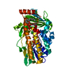
| Title | Crystal structure of a gdsl-like lipase (alr1529) from nostoc sp. pcc 7120 at 2.01 A resolution | ||||||
 Components Components | putative lipase from the G-D-S-L family | ||||||
 Keywords Keywords | HYDROLASE / Putative lipase from the g-d-s-l family / structural genomics / Joint Center for Structural Genomics / JCSG / Protein Structure Initiative / PSI | ||||||
| Function / homology | Isoamyl acetate-hydrolyzing esterase Iah1-like / SGNH hydrolase / SGNH hydrolase-type esterase domain / GDSL-like Lipase/Acylhydrolase family / SGNH hydrolase superfamily / Rossmann fold / 3-Layer(aba) Sandwich / Alpha Beta / Alr1529 protein Function and homology information Function and homology information | ||||||
| Biological species |  Nostoc sp. (bacteria) Nostoc sp. (bacteria) | ||||||
| Method |  X-RAY DIFFRACTION / SYNCHROTRON / MOLECULAR REPLACEMENT / Resolution: 2.01 Å X-RAY DIFFRACTION / SYNCHROTRON / MOLECULAR REPLACEMENT / Resolution: 2.01 Å | ||||||
 Authors Authors | Joint Center for Structural Genomics (JCSG) | ||||||
 Citation Citation | Journal: To be published Title: Crystal structure of putative lipase from the G-D-S-L family from Nostoc sp. at 2.01 A resolution Authors: Joint Center for Structural Genomics (JCSG) | ||||||
| History |
|
- Structure visualization
| Structure viewer | Molecule: MolmilJmol/JSmol |
|---|
- Downloads & links
-Download
| PDBx/mmCIF format | 1vjg.cif.gz | 56.8 KB | Display | PDBx/mmCIF format |
|---|---|---|---|---|
| PDB format | pdb1vjg.ent.gz | 40.4 KB | Display | PDB format |
| PDBx/mmJSON format | 1vjg.json.gz | Tree view | PDBx/mmJSON format | |
| Others |  Other downloads Other downloads |
-Validation report
| Arichive directory | https://data.pdbj.org/pub/pdb/validation_reports/vj/1vjgftp://data.pdbj.org/pub/pdb/validation_reports/vj/1vjg | HTTPS FTP |
|---|
-Related structure data
| Similar structure data | |
|---|---|
| Other databases |


-Links
 PDBj
PDBj- Assembly
| Deposited unit | 
| ||||||||
|---|---|---|---|---|---|---|---|---|---|
| 1 | 
| ||||||||
| Unit cell |
|
-Components
| #1: Protein | Mass: 25133.396 Da / Num. of mol.: 1 Source method: isolated from a genetically manipulated source Source: (gene. exp.) Nostoc sp. (bacteria) / Strain: PCC 7120 / Gene: ALR1529 / Production host: |
|---|---|
| #2: Water | ChemComp-HOH /  Mass: 18.015 Da / Num. of mol.: 92 / Source method: isolated from a natural source / Formula: H2O Mass: 18.015 Da / Num. of mol.: 92 / Source method: isolated from a natural source / Formula: H2O |
| Has protein modification | Y |
-Experimental details
-Experiment
| Experiment | Method: X-RAY DIFFRACTION / Number of used crystals: 1 |
|---|
- Sample preparation
| Crystal | Density Matthews: 2.43 Å3/Da / Density % sol: 48.97 % |
|---|---|
| Crystal grow | Temperature: 277 K / Method: vapor diffusion, sitting drop, nanodrop / pH: 7.5 Details: 10% iso-propanol, 20% PEG-4000, 0.1M Sodium HEPES pH 7.5, VAPOR DIFFUSION,SITTING DROP,NANODROP, temperature 277K |
-Data collection
| Diffraction | Mean temperature: 100 K |
|---|---|
| Diffraction source | Source: SYNCHROTRON / Site: ALS  / Beamline: 8.2.1 / Wavelength: 0.9801 / Beamline: 8.2.1 / Wavelength: 0.9801 |
| Detector | Type: ADSC / Detector: CCD / Date: Jul 13, 2003 |
| Radiation | Monochromator: Double Crystal Si(111) / Protocol: SINGLE WAVELENGTH / Monochromatic (M) / Laue (L): M / Scattering type: x-ray |
| Radiation wavelength | Wavelength: 0.9801 Å / Relative weight: 1 |
| Reflection | Resolution: 2.01→43.32 Å / Num. obs: 15039 / % possible obs: 90.6 % / Redundancy: 5.6 % / Biso Wilson estimate: 57.4 Å2 / Rsym value: 0.07 / Net I/σ(I): 13 |
| Reflection shell | Resolution: 2.01→2.12 Å / Redundancy: 2.8 % / Mean I/σ(I) obs: 3.4 / Num. unique all: 1526 / Rsym value: 0.277 / % possible all: 65.7 |
- Processing
| Software |
| ||||||||||||||||||||||||||||||||||||||||||||||||||||||||||||||||||||||||||||||||||||||||||||||||||||
|---|---|---|---|---|---|---|---|---|---|---|---|---|---|---|---|---|---|---|---|---|---|---|---|---|---|---|---|---|---|---|---|---|---|---|---|---|---|---|---|---|---|---|---|---|---|---|---|---|---|---|---|---|---|---|---|---|---|---|---|---|---|---|---|---|---|---|---|---|---|---|---|---|---|---|---|---|---|---|---|---|---|---|---|---|---|---|---|---|---|---|---|---|---|---|---|---|---|---|---|---|---|
| Refinement | Method to determine structure: MOLECULAR REPLACEMENT / Resolution: 2.01→43.32 Å / Cor.coef. Fo:Fc: 0.968 / Cor.coef. Fo:Fc free: 0.953 / SU B: 3.678 / SU ML: 0.1 / TLS residual ADP flag: LIKELY RESIDUAL / Cross valid method: THROUGHOUT / ESU R: 0.177 / ESU R Free: 0.161 / Details: HYDROGENS HAVE BEEN ADDED IN THE RIDING POSITIONS
| ||||||||||||||||||||||||||||||||||||||||||||||||||||||||||||||||||||||||||||||||||||||||||||||||||||
| Solvent computation | Ion probe radii: 0.8 Å / Shrinkage radii: 0.8 Å / VDW probe radii: 1.4 Å / Solvent model: BABINET MODEL WITH MASK | ||||||||||||||||||||||||||||||||||||||||||||||||||||||||||||||||||||||||||||||||||||||||||||||||||||
| Displacement parameters | Biso mean: 41.592 Å2
| ||||||||||||||||||||||||||||||||||||||||||||||||||||||||||||||||||||||||||||||||||||||||||||||||||||
| Refinement step | Cycle: LAST / Resolution: 2.01→43.32 Å
| ||||||||||||||||||||||||||||||||||||||||||||||||||||||||||||||||||||||||||||||||||||||||||||||||||||
| Refine LS restraints |
| ||||||||||||||||||||||||||||||||||||||||||||||||||||||||||||||||||||||||||||||||||||||||||||||||||||
| LS refinement shell | Resolution: 2.01→2.061 Å / Total num. of bins used: 20
| ||||||||||||||||||||||||||||||||||||||||||||||||||||||||||||||||||||||||||||||||||||||||||||||||||||
| Refinement TLS params. | Method: refined / Origin x: 1.66 Å / Origin y: 36.085 Å / Origin z: 16.352 Å
| ||||||||||||||||||||||||||||||||||||||||||||||||||||||||||||||||||||||||||||||||||||||||||||||||||||
| Refinement TLS group | Selection: ALL |
