 Movie
Movie Controller


+ Open data

- Basic information
| Entry | Database: PDB / ID: 1vio | ||||||
|---|---|---|---|---|---|---|---|
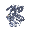
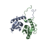
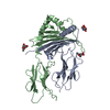
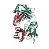
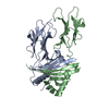
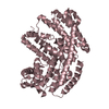
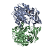
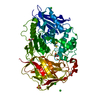
| Title | Crystal structure of pseudouridylate synthase | ||||||
 Components Components | Ribosomal small subunit pseudouridine synthase A | ||||||
 Keywords Keywords | LYASE / structural genomics | ||||||
| Function / homology |  Function and homology information Function and homology information16S rRNA pseudouridine516 synthase / 16S rRNA pseudouridine(516) synthase activity / enzyme-directed rRNA pseudouridine synthesis / pseudouridine synthase activity / RNA binding / cytosol Similarity search - Function | ||||||
| Biological species |  Haemophilus influenzae (bacteria) Haemophilus influenzae (bacteria) | ||||||
| Method |  X-RAY DIFFRACTION / SYNCHROTRON / MOLECULAR REPLACEMENT / Resolution: 1.59 Å X-RAY DIFFRACTION / SYNCHROTRON / MOLECULAR REPLACEMENT / Resolution: 1.59 Å | ||||||
 Authors Authors | Structural GenomiX | ||||||
 Citation Citation | Journal: Acta Crystallogr.,Sect.F / Year: 2005 Title: Structure of the pseudouridine synthase RsuA from Haemophilus influenzae. Authors: Matte, A. / Louie, G.V. / Sivaraman, J. / Cygler, M. / Burley, S.K. #1: Journal: Proteins / Year: 2005Title: Structural analysis of a set of proteins resulting from a bacterial genomics project Authors: Badger, J. / Sauder, J.M. / Adams, J.M. / Antonysamy, S. / Bain, K. / Bergseid, M.G. / Buchanan, S.G. / Buchanan, M.D. / Batiyenko, Y. / Christopher, J.A. / Emtage, S. / Eroshkina, A. / ...Authors: Badger, J. / Sauder, J.M. / Adams, J.M. / Antonysamy, S. / Bain, K. / Bergseid, M.G. / Buchanan, S.G. / Buchanan, M.D. / Batiyenko, Y. / Christopher, J.A. / Emtage, S. / Eroshkina, A. / Feil, I. / Furlong, E.B. / Gajiwala, K.S. / Gao, X. / He, D. / Hendle, J. / Huber, A. / Hoda, K. / Kearins, P. / Kissinger, C. / Laubert, B. / Lewis, H.A. / Lin, J. / Loomis, K. / Lorimer, D. / Louie, G. / Maletic, M. / Marsh, C.D. / Miller, I. / Molinari, J. / Muller-Dieckmann, H.J. / Newman, J.M. / Noland, B.W. / Pagarigan, B. / Park, F. / Peat, T.S. / Post, K.W. / Radojicic, S. / Ramos, A. / Romero, R. / Rutter, M.E. / Sanderson, W.E. / Schwinn, K.D. / Tresser, J. / Winhoven, J. / Wright, T.A. / Wu, L. / Xu, J. / Harris, T.J. | ||||||
| History |
|
- Structure visualization
| Structure viewer | Molecule: MolmilJmol/JSmol |
|---|
- Downloads & links
-Download
| PDBx/mmCIF format | 1vio.cif.gz | 115 KB | Display | PDBx/mmCIF format |
|---|---|---|---|---|
| PDB format | pdb1vio.ent.gz | 88.4 KB | Display | PDB format |
| PDBx/mmJSON format | 1vio.json.gz | Tree view | PDBx/mmJSON format | |
| Others |  Other downloads Other downloads |
-Validation report
| Arichive directory | https://data.pdbj.org/pub/pdb/validation_reports/vi/1vioftp://data.pdbj.org/pub/pdb/validation_reports/vi/1vio | HTTPS FTP |
|---|
-Related structure data
| Similar structure data |
|---|


-Links
 PDBj
PDBj


- Assembly
| Deposited unit | 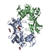
| ||||||||
|---|---|---|---|---|---|---|---|---|---|
| 1 |
| ||||||||
| Unit cell |
|
-Components
| #1: Protein | Mass: 27474.822 Da / Num. of mol.: 2 Source method: isolated from a genetically manipulated source Source: (gene. exp.) Haemophilus influenzae (bacteria) / Gene: RSUA, HI1243 / Production host: #2: Chemical | ChemComp-BU1 /   Mass: 90.121 Da / Num. of mol.: 4 / Source method: obtained synthetically / Formula: C4H10O2 Mass: 90.121 Da / Num. of mol.: 4 / Source method: obtained synthetically / Formula: C4H10O2#3: Water | ChemComp-HOH / |  Mass: 18.015 Da / Num. of mol.: 528 / Source method: isolated from a natural source / Formula: H2O Mass: 18.015 Da / Num. of mol.: 528 / Source method: isolated from a natural source / Formula: H2O |
|---|
-Experimental details
-Experiment
| Experiment | Method: X-RAY DIFFRACTION / Number of used crystals: 1 |
|---|
- Sample preparation
| Crystal | Density Matthews: 2.38 Å3/Da / Density % sol: 48.31 % | |||||||||||||||||||||||||||||||||||||||||||||||||||||||||||||||
|---|---|---|---|---|---|---|---|---|---|---|---|---|---|---|---|---|---|---|---|---|---|---|---|---|---|---|---|---|---|---|---|---|---|---|---|---|---|---|---|---|---|---|---|---|---|---|---|---|---|---|---|---|---|---|---|---|---|---|---|---|---|---|---|---|
| Crystal grow | *PLUS Temperature: 277 K / pH: 7.5 / Method: vapor diffusion, hanging drop | |||||||||||||||||||||||||||||||||||||||||||||||||||||||||||||||
| Components of the solutions | *PLUS
|
-Data collection
| Diffraction source | Source: SYNCHROTRON / Site: APS  / Beamline: 32-ID / Wavelength: 0.9793 Å / Beamline: 32-ID / Wavelength: 0.9793 Å |
|---|---|
| Detector | Type: MARRESEARCH / Detector: CCD |
| Radiation | Protocol: SINGLE WAVELENGTH / Monochromatic (M) / Laue (L): M / Scattering type: x-ray |
| Radiation wavelength | Wavelength: 0.9793 Å / Relative weight: 1 |
| Reflection | Resolution: 1.51→22.14 Å / Num. all: 80022 / Num. obs: 80022 / % possible obs: 99.6 % / Redundancy: 7.3 % / Rmerge(I) obs: 0.087 / Net I/σ(I): 11.5 |
| Reflection shell | Resolution: 1.51→1.59 Å / Redundancy: 6.2 % / Rmerge(I) obs: 0.01109 / Mean I/σ(I) obs: 1.8 / % possible all: 97.4 |
| Reflection | *PLUS Lowest resolution: 22 Å / Num. obs: 80195 / Num. measured all: 587441 |
| Reflection shell | *PLUS % possible obs: 97.4 % |
- Processing
| Software |
| ||||||||||||||||||||||||||||||
|---|---|---|---|---|---|---|---|---|---|---|---|---|---|---|---|---|---|---|---|---|---|---|---|---|---|---|---|---|---|---|---|
| Refinement | Method to determine structure: MOLECULAR REPLACEMENT / Resolution: 1.59→22.14 Å / σ(F): 0
| ||||||||||||||||||||||||||||||
| Solvent computation | Solvent model: Babinet bulk solvent correction / Bsol: 165.646 Å2 / ksol: 0.783 e/Å3 | ||||||||||||||||||||||||||||||
| Displacement parameters | Biso mean: 21.208 Å2
| ||||||||||||||||||||||||||||||
| Refine Biso | Class: all / Treatment: isotropic | ||||||||||||||||||||||||||||||
| Refinement step | Cycle: LAST / Resolution: 1.59→22.14 Å
| ||||||||||||||||||||||||||||||
| Refine LS restraints |
| ||||||||||||||||||||||||||||||
| Software | *PLUS Version: 4 / Classification: refinement | ||||||||||||||||||||||||||||||
| Refinement | *PLUS Lowest resolution: 22.1 Å | ||||||||||||||||||||||||||||||
| Solvent computation | *PLUS | ||||||||||||||||||||||||||||||
| Displacement parameters | *PLUS | ||||||||||||||||||||||||||||||
| Refine LS restraints | *PLUS Type: p_angle_d / Dev ideal: 1.45 | ||||||||||||||||||||||||||||||
| LS refinement shell | *PLUS Num. reflection Rfree: 3437 / Num. reflection Rwork: 65715 |
