 Movie
Movie Controller


+ Open data

- Basic information
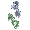
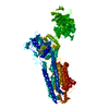
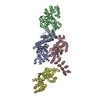
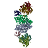
| Entry | Database: PDB / ID: 1vfp | ||||||
|---|---|---|---|---|---|---|---|
| Title | Crystal structure of the SR CA2+-ATPase with bound AMPPCP | ||||||
 Components Components | Sarcoplasmic/endoplasmic reticulum calcium ATPase 1 | ||||||
 Keywords Keywords | HYDROLASE / Membrane protein / P-type ATPase / HAD fold | ||||||
| Function / homology |  Function and homology information Function and homology informationpositive regulation of cardiac muscle cell contraction / positive regulation of calcium ion import into sarcoplasmic reticulum / positive regulation of ATPase-coupled calcium transmembrane transporter activity / positive regulation of fast-twitch skeletal muscle fiber contraction / H zone / regulation of striated muscle contraction / calcium ion import into sarcoplasmic reticulum / negative regulation of striated muscle contraction / P-type Ca2+ transporter / P-type calcium transporter activity ...positive regulation of cardiac muscle cell contraction / positive regulation of calcium ion import into sarcoplasmic reticulum / positive regulation of ATPase-coupled calcium transmembrane transporter activity / positive regulation of fast-twitch skeletal muscle fiber contraction / H zone / regulation of striated muscle contraction / calcium ion import into sarcoplasmic reticulum / negative regulation of striated muscle contraction / P-type Ca2+ transporter / P-type calcium transporter activity / I band / endoplasmic reticulum-Golgi intermediate compartment / sarcoplasmic reticulum membrane / sarcoplasmic reticulum / calcium ion transport / calcium ion binding / endoplasmic reticulum membrane / perinuclear region of cytoplasm / endoplasmic reticulum / ATP hydrolysis activity / ATP binding / membrane Similarity search - Function | ||||||
| Biological species |  | ||||||
| Method |  X-RAY DIFFRACTION / SYNCHROTRON / MOLECULAR REPLACEMENT / Resolution: 2.9 Å X-RAY DIFFRACTION / SYNCHROTRON / MOLECULAR REPLACEMENT / Resolution: 2.9 Å | ||||||
 Authors Authors | Toyoshima, C. / Mizutani, T. | ||||||
 Citation Citation | Journal: Nature / Year: 2004 Title: Crystal structure of the calcium pump with a bound ATP analogue. Authors: Toyoshima, C. / Mizutani, T. #1: Journal: Nature / Year: 2000 Title: Crystal structure of the calcium pump of sarcoplasmic reticulum at 2.6 A resolution. Authors: Toyoshima, C. / Nakasako, M. / Nomura, H. / Ogawa, H. #2: Journal: Nature / Year: 2002 Title: Structural changes in the calcium pump accompanying the dissociation of calcium. Authors: Toyoshima, C. / Nomura, H. #3: Journal: FEBS Lett. / Year: 2003 Title: Structural basis of ion pumping by Ca(2+)-ATPase of sarcoplasmic reticulum. Authors: Toyoshima, C. / Nomura, H. / Sugita, Y. | ||||||
| History |
|
- Structure visualization
| Structure viewer | Molecule: MolmilJmol/JSmol |
|---|
- Downloads & links
-Download
| PDBx/mmCIF format | 1vfp.cif.gz | 358 KB | Display | PDBx/mmCIF format |
|---|---|---|---|---|
| PDB format | pdb1vfp.ent.gz | 292.9 KB | Display | PDB format |
| PDBx/mmJSON format | 1vfp.json.gz | Tree view | PDBx/mmJSON format | |
| Others |  Other downloads Other downloads |
-Validation report
| Summary document | 1vfp_validation.pdf.gz | 513.1 KB | Display | wwPDB validaton report |
|---|---|---|---|---|
| Full document | 1vfp_full_validation.pdf.gz | 588.3 KB | Display | |
| Data in XML | 1vfp_validation.xml.gz | 48.6 KB | Display | |
| Data in CIF | 1vfp_validation.cif.gz | 69.9 KB | Display | |
| Arichive directory | https://data.pdbj.org/pub/pdb/validation_reports/vf/1vfpftp://data.pdbj.org/pub/pdb/validation_reports/vf/1vfp | HTTPS FTP |
-Related structure data
| Related structure data |  1eul S: Starting model for refinement |
|---|---|
| Similar structure data |


-Links
 PDBj
PDBj


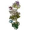
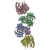
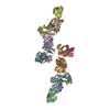
- Assembly
| Deposited unit | 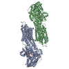
| ||||||||
|---|---|---|---|---|---|---|---|---|---|
| 1 |
| ||||||||
| Unit cell |
| ||||||||
| Noncrystallographic symmetry (NCS) | NCS oper: (Code: given Matrix: (-0.65592, 0.74829, 0.09915), Vector: |
-Components
| #1: Protein | Mass: 109602.578 Da / Num. of mol.: 2 / Source method: isolated from a natural source / Source: (natural) #2: Chemical | ChemComp-CA /   Mass: 40.078 Da / Num. of mol.: 4 / Source method: obtained synthetically / Formula: Ca Mass: 40.078 Da / Num. of mol.: 4 / Source method: obtained synthetically / Formula: Ca#3: Chemical |   Mass: 24.305 Da / Num. of mol.: 2 / Source method: obtained synthetically / Formula: Mg Mass: 24.305 Da / Num. of mol.: 2 / Source method: obtained synthetically / Formula: Mg#4: Chemical |   Mass: 505.208 Da / Num. of mol.: 2 / Source method: obtained synthetically / Formula: C11H18N5O12P3 / Comment: AMP-PCP, energy-carrying molecule analogue*YM Mass: 505.208 Da / Num. of mol.: 2 / Source method: obtained synthetically / Formula: C11H18N5O12P3 / Comment: AMP-PCP, energy-carrying molecule analogue*YM#5: Water | ChemComp-HOH / |  Mass: 18.015 Da / Num. of mol.: 8 / Source method: isolated from a natural source / Formula: H2O Mass: 18.015 Da / Num. of mol.: 8 / Source method: isolated from a natural source / Formula: H2OHas protein modification | Y | |
|---|
-Experimental details
-Experiment
| Experiment | Method: X-RAY DIFFRACTION / Number of used crystals: 2 |
|---|
- Sample preparation
| Crystal | Density Matthews: 3.714 Å3/Da / Density % sol: 68.2 % | ||||||||||||||||||||||||||||||||||||||||||||||||||||||||||||
|---|---|---|---|---|---|---|---|---|---|---|---|---|---|---|---|---|---|---|---|---|---|---|---|---|---|---|---|---|---|---|---|---|---|---|---|---|---|---|---|---|---|---|---|---|---|---|---|---|---|---|---|---|---|---|---|---|---|---|---|---|---|
| Crystal grow | Method: microdialysis / pH: 6.1 / Details: PEG400, pH 6.1, MICRODIALYSIS | ||||||||||||||||||||||||||||||||||||||||||||||||||||||||||||
| Crystal grow | *PLUS | ||||||||||||||||||||||||||||||||||||||||||||||||||||||||||||
| Components of the solutions | *PLUS
|
-Data collection
| Diffraction | Mean temperature: 100 K |
|---|---|
| Diffraction source | Source: SYNCHROTRON / Site: SPring-8  / Beamline: BL41XU / Wavelength: 0.72 Å / Beamline: BL41XU / Wavelength: 0.72 Å |
| Detector | Type: MARRESEARCH / Detector: CCD / Date: Apr 9, 2002 |
| Radiation | Monochromator: rotated-inclined double-crystal / Protocol: SINGLE WAVELENGTH / Monochromatic (M) / Laue (L): M / Scattering type: x-ray |
| Radiation wavelength | Wavelength: 0.72 Å / Relative weight: 1 |
| Reflection | Resolution: 2.9→20 Å / Num. all: 70956 / Num. obs: 70956 / % possible obs: 99 % / Observed criterion σ(I): -3 / Redundancy: 6.37 % / Biso Wilson estimate: 63.3 Å2 / Rmerge(I) obs: 0.056 / Net I/σ(I): 26.45 |
| Reflection shell | Resolution: 2.9→2.98 Å / Redundancy: 3.1 % / Rmerge(I) obs: 0.24 / Mean I/σ(I) obs: 3.598 / Num. unique all: 5239 / % possible all: 94.2 |
| Reflection | *PLUS Lowest resolution: 20 Å |
- Processing
| Software |
| ||||||||||||||||||||||||||||||||||||||||||||||||||||||||||||
|---|---|---|---|---|---|---|---|---|---|---|---|---|---|---|---|---|---|---|---|---|---|---|---|---|---|---|---|---|---|---|---|---|---|---|---|---|---|---|---|---|---|---|---|---|---|---|---|---|---|---|---|---|---|---|---|---|---|---|---|---|---|
| Refinement | Method to determine structure: MOLECULAR REPLACEMENT Starting model: P-domain and M3-M10 transmembrane helices of 1EUL 1eul Resolution: 2.9→15 Å / Rfactor Rfree error: 0.005 / Data cutoff high absF: 232616.95 / Data cutoff low absF: 0 / Isotropic thermal model: GROUP / Cross valid method: THROUGHOUT / σ(F): 2 / Stereochemistry target values: Engh & Huber
| ||||||||||||||||||||||||||||||||||||||||||||||||||||||||||||
| Solvent computation | Solvent model: FLAT MODEL / Bsol: 34.83 Å2 / ksol: 0.28476 e/Å3 | ||||||||||||||||||||||||||||||||||||||||||||||||||||||||||||
| Displacement parameters | Biso mean: 75.3 Å2
| ||||||||||||||||||||||||||||||||||||||||||||||||||||||||||||
| Refine analyze |
| ||||||||||||||||||||||||||||||||||||||||||||||||||||||||||||
| Refinement step | Cycle: LAST / Resolution: 2.9→15 Å
| ||||||||||||||||||||||||||||||||||||||||||||||||||||||||||||
| Refine LS restraints |
| ||||||||||||||||||||||||||||||||||||||||||||||||||||||||||||
| Refine LS restraints NCS | NCS model details: CONSTR | ||||||||||||||||||||||||||||||||||||||||||||||||||||||||||||
| LS refinement shell | Resolution: 2.9→3.08 Å / Rfactor Rfree error: 0.015 / Total num. of bins used: 6
| ||||||||||||||||||||||||||||||||||||||||||||||||||||||||||||
| Xplor file |
| ||||||||||||||||||||||||||||||||||||||||||||||||||||||||||||
| Software | *PLUS Version: 1.1 / Classification: refinement | ||||||||||||||||||||||||||||||||||||||||||||||||||||||||||||
| Refinement | *PLUS σ(F): 2 / % reflection Rfree: 5.1 % / Rfactor obs: 0.252 / Rfactor Rfree: 0.296 | ||||||||||||||||||||||||||||||||||||||||||||||||||||||||||||
| Solvent computation | *PLUS | ||||||||||||||||||||||||||||||||||||||||||||||||||||||||||||
| Displacement parameters | *PLUS | ||||||||||||||||||||||||||||||||||||||||||||||||||||||||||||
| Refine LS restraints | *PLUS
| ||||||||||||||||||||||||||||||||||||||||||||||||||||||||||||
| LS refinement shell | *PLUS Rfactor Rfree: 0.365 / % reflection Rfree: 5.3 % / Rfactor Rwork: 0.34 |
