 Movie
Movie Controller

[English] 日本語
 Yorodumi
- PDB-1vev: Crystal structure of peptide deformylase from Leptospira Interrog... -
+ Open data

- Basic information
| Entry | Database: PDB / ID: 1vev | ||||||
|---|---|---|---|---|---|---|---|
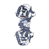
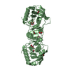
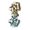
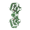
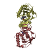
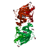
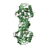
| Title | Crystal structure of peptide deformylase from Leptospira Interrogans (LiPDF) at pH6.5 | ||||||
 Components Components | Peptide deformylase | ||||||
 Keywords Keywords | HYDROLASE / closed conformation / MES | ||||||
| Function / homology |  Function and homology information Function and homology informationpeptide deformylase / peptide deformylase activity / translation / metal ion binding Similarity search - Function | ||||||
| Biological species |  Leptospira interrogans (bacteria) Leptospira interrogans (bacteria) | ||||||
| Method |  X-RAY DIFFRACTION / SYNCHROTRON / MOLECULAR REPLACEMENT / Resolution: 2.51 Å X-RAY DIFFRACTION / SYNCHROTRON / MOLECULAR REPLACEMENT / Resolution: 2.51 Å | ||||||
 Authors Authors | Zhou, Z. / Song, X. / Li, Y. / Gong, W. | ||||||
 Citation Citation | Journal: J.Biol.Chem. / Year: 2005 Title: Novel conformational states of peptide deformylase from pathogenic bacterium Leptospira interrogans: implications for population shift Authors: Zhou, Z. / Song, X. / Gong, W. | ||||||
| History |
|
- Structure visualization
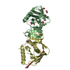
| Structure viewer | Molecule: MolmilJmol/JSmol |
|---|
- Downloads & links
-Download
| PDBx/mmCIF format | 1vev.cif.gz | 85.4 KB | Display | PDBx/mmCIF format |
|---|---|---|---|---|
| PDB format | pdb1vev.ent.gz | 63.8 KB | Display | PDB format |
| PDBx/mmJSON format | 1vev.json.gz | Tree view | PDBx/mmJSON format | |
| Others |  Other downloads Other downloads |
-Validation report
| Arichive directory | https://data.pdbj.org/pub/pdb/validation_reports/ve/1vevftp://data.pdbj.org/pub/pdb/validation_reports/ve/1vev | HTTPS FTP |
|---|
-Related structure data
| Related structure data |  1sv2SC  1szzC  1veyC  1vezC S: Starting model for refinement C: citing same article ( |
|---|---|
| Similar structure data |


-Links
 PDBj
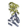
PDBj- Assembly
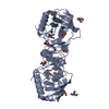
| Deposited unit | 
| ||||||||
|---|---|---|---|---|---|---|---|---|---|
| 1 | 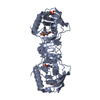
| ||||||||
| 2 | 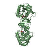
| ||||||||
| Unit cell |
| ||||||||
| Details | The biological unit 1 is generated by chain A (x,y,z (1_555),1-y,1-x,1/2-z(8_665)). / The biological unit 2 is generated by chain B (x,y,z (1_555),1+y,-1+x,-z(7_645)). |
-Components
| #1: Protein | Mass: 20274.117 Da / Num. of mol.: 2 Source method: isolated from a genetically manipulated source Source: (gene. exp.) Leptospira interrogans (bacteria) / Plasmid: pET22b / Species (production host): Escherichia coli / Production host: #2: Chemical |   Mass: 65.409 Da / Num. of mol.: 2 / Source method: obtained synthetically / Formula: Zn Mass: 65.409 Da / Num. of mol.: 2 / Source method: obtained synthetically / Formula: Zn#3: Chemical |   Mass: 195.237 Da / Num. of mol.: 3 / Source method: obtained synthetically / Formula: C6H13NO4S / Comment: pH buffer*YM Mass: 195.237 Da / Num. of mol.: 3 / Source method: obtained synthetically / Formula: C6H13NO4S / Comment: pH buffer*YM#4: Chemical |   Mass: 46.025 Da / Num. of mol.: 2 / Source method: obtained synthetically / Formula: CH2O2 Mass: 46.025 Da / Num. of mol.: 2 / Source method: obtained synthetically / Formula: CH2O2#5: Water | ChemComp-HOH / |  Mass: 18.015 Da / Num. of mol.: 210 / Source method: isolated from a natural source / Formula: H2O Mass: 18.015 Da / Num. of mol.: 210 / Source method: isolated from a natural source / Formula: H2O |
|---|
-Experimental details
-Experiment
| Experiment | Method: X-RAY DIFFRACTION / Number of used crystals: 1 |
|---|
- Sample preparation
| Crystal | Density Matthews: 4.34 Å3/Da / Density % sol: 70 % |
|---|---|
| Crystal grow | Temperature: 277 K / Method: vapor diffusion, hanging drop / pH: 6.5 Details: 4M formate sodium, pH 6.5, VAPOR DIFFUSION, HANGING DROP, temperature 277K |
-Data collection
| Diffraction | Mean temperature: 100 K |
|---|---|
| Diffraction source | Source: SYNCHROTRON / Site: BSRF  / Beamline: 3W1A / Wavelength: 1.2 Å / Beamline: 3W1A / Wavelength: 1.2 Å |
| Detector | Type: MARRESEARCH / Detector: CCD / Date: Mar 1, 2003 |
| Radiation | Protocol: SINGLE WAVELENGTH / Monochromatic (M) / Laue (L): M / Scattering type: x-ray |
| Radiation wavelength | Wavelength: 1.2 Å / Relative weight: 1 |
| Reflection | Resolution: 2.51→43.91 Å / Num. all: 24689 / Num. obs: 21663 / % possible obs: 87.7 % / Observed criterion σ(F): 0 / Observed criterion σ(I): 0 |
| Reflection shell | Resolution: 2.5→2.66 Å / % possible all: 75.9 |
- Processing
| Software |
| |||||||||||||||||||||||||
|---|---|---|---|---|---|---|---|---|---|---|---|---|---|---|---|---|---|---|---|---|---|---|---|---|---|---|
| Refinement | Method to determine structure: MOLECULAR REPLACEMENT Starting model: PDB id 1SV2 Resolution: 2.51→43.91 Å / Cross valid method: THROUGHOUT / σ(F): 0 / Stereochemistry target values: Engh & Huber
| |||||||||||||||||||||||||
| Refine analyze |
| |||||||||||||||||||||||||
| Refinement step | Cycle: LAST / Resolution: 2.51→43.91 Å
| |||||||||||||||||||||||||
| Refine LS restraints |
|
