 Movie
Movie Controller

[English] 日本語
 Yorodumi
- PDB-1vd6: Crystal Structure of Glycerophosphoryl Diester Phosphodiesterase ... -
+ Open data

- Basic information
| Entry | Database: PDB / ID: 1vd6 | ||||||
|---|---|---|---|---|---|---|---|
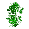
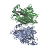
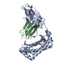
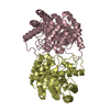
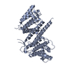
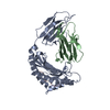
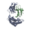
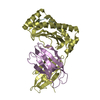
| Title | Crystal Structure of Glycerophosphoryl Diester Phosphodiesterase complexed with Glycerol | ||||||
 Components Components | Glycerophosphoryl Diester Phosphodiesterase | ||||||
 Keywords Keywords | HYDROLASE / Glycerophosphoryl Diester Phosphodiesterase / Glycerophosphodiester Phosphodiesterase / Thermus thermophilus / HB8 / Glycerol Complex / RIKEN Structural Genomics/Proteomics Initiative / RSGI / Structural Genomics | ||||||
| Function / homology |  Function and homology information Function and homology information | ||||||
| Biological species |   Thermus thermophilus (bacteria) Thermus thermophilus (bacteria) | ||||||
| Method |  X-RAY DIFFRACTION / SYNCHROTRON / MOLECULAR REPLACEMENT / Resolution: 1.3 Å X-RAY DIFFRACTION / SYNCHROTRON / MOLECULAR REPLACEMENT / Resolution: 1.3 Å | ||||||
 Authors Authors | Ishijima, J. / Yutani, K. / RIKEN Structural Genomics/Proteomics Initiative (RSGI) | ||||||
 Citation Citation | Journal: To be Published Title: Crystal Structure of Glycerophosphoryl Diester Phosphodiesterase complexed with Glycerol Authors: Ishijima, J. / Yutani, K. | ||||||
| History |
|
- Structure visualization
| Structure viewer | Molecule: MolmilJmol/JSmol |
|---|
- Downloads & links
-Download
| PDBx/mmCIF format | 1vd6.cif.gz | 62.7 KB | Display | PDBx/mmCIF format |
|---|---|---|---|---|
| PDB format | pdb1vd6.ent.gz | 45.7 KB | Display | PDB format |
| PDBx/mmJSON format | 1vd6.json.gz | Tree view | PDBx/mmJSON format | |
| Others |  Other downloads Other downloads |
-Validation report
| Summary document | 1vd6_validation.pdf.gz | 439.8 KB | Display | wwPDB validaton report |
|---|---|---|---|---|
| Full document | 1vd6_full_validation.pdf.gz | 442.7 KB | Display | |
| Data in XML | 1vd6_validation.xml.gz | 13.9 KB | Display | |
| Data in CIF | 1vd6_validation.cif.gz | 21 KB | Display | |
| Arichive directory | https://data.pdbj.org/pub/pdb/validation_reports/vd/1vd6ftp://data.pdbj.org/pub/pdb/validation_reports/vd/1vd6 | HTTPS FTP |
-Related structure data
| Related structure data |  1v8eS S: Starting model for refinement |
|---|---|
| Similar structure data | |
| Other databases |


-Links
 PDBj
PDBj- Assembly
| Deposited unit | 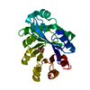
| ||||||||
|---|---|---|---|---|---|---|---|---|---|
| 1 | 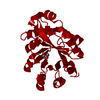
| ||||||||
| Unit cell |
| ||||||||
| Details | The biological assembly is a dimer covalently bound by disulfide bond. |
-Components
| #1: Protein | Mass: 24435.143 Da / Num. of mol.: 1 Source method: isolated from a genetically manipulated source Source: (gene. exp.) Thermus thermophilus (bacteria) / Strain: HB8 / Plasmid: pET11a / Production host: References: GenBank: 55978324, UniProt: Q53W25*PLUS, glycerophosphodiester phosphodiesterase |
|---|---|
| #2: Chemical | ChemComp-GOL /   Mass: 92.094 Da / Num. of mol.: 1 / Source method: obtained synthetically / Formula: C3H8O3 Mass: 92.094 Da / Num. of mol.: 1 / Source method: obtained synthetically / Formula: C3H8O3 |
| #3: Water | ChemComp-HOH /  Mass: 18.015 Da / Num. of mol.: 317 / Source method: isolated from a natural source / Formula: H2O Mass: 18.015 Da / Num. of mol.: 317 / Source method: isolated from a natural source / Formula: H2O |
| Has protein modification | Y |
-Experimental details
-Experiment
| Experiment | Method: X-RAY DIFFRACTION / Number of used crystals: 1 |
|---|
- Sample preparation
| Crystal | Density Matthews: 2.05 Å3/Da / Density % sol: 39.65 % |
|---|---|
| Crystal grow | Temperature: 291 K / Method: microbatch / pH: 8.1 Details: Ammonium sulfate, dioxane, pH 8.1, Microbatch, temperature 291K |
-Data collection
| Diffraction | Mean temperature: 100 K |
|---|---|
| Diffraction source | Source: SYNCHROTRON / Site: SPring-8  / Beamline: BL26B1 / Wavelength: 1 Å / Beamline: BL26B1 / Wavelength: 1 Å |
| Detector | Type: RIGAKU RAXIS V / Detector: IMAGE PLATE / Date: Jun 24, 2003 |
| Radiation | Monochromator: mirror / Protocol: SINGLE WAVELENGTH / Monochromatic (M) / Laue (L): M / Scattering type: x-ray |
| Radiation wavelength | Wavelength: 1 Å / Relative weight: 1 |
| Reflection | Resolution: 1.3→50 Å / Num. all: 55698 / Num. obs: 55698 / % possible obs: 96.5 % / Observed criterion σ(F): 0 / Observed criterion σ(I): 0 / Redundancy: 10.5 % / Biso Wilson estimate: 15.4 Å2 / Rmerge(I) obs: 0.074 / Net I/σ(I): 19 |
| Reflection shell | Resolution: 1.3→1.35 Å / Redundancy: 9.9 % / Rmerge(I) obs: 0.478 / Mean I/σ(I) obs: 4 / Num. unique all: 5701 / % possible all: 100 |
- Processing
| Software |
| ||||||||||||||||||||||||||||||||||||||||||||||||||||||||||||||||||||||||||||||||
|---|---|---|---|---|---|---|---|---|---|---|---|---|---|---|---|---|---|---|---|---|---|---|---|---|---|---|---|---|---|---|---|---|---|---|---|---|---|---|---|---|---|---|---|---|---|---|---|---|---|---|---|---|---|---|---|---|---|---|---|---|---|---|---|---|---|---|---|---|---|---|---|---|---|---|---|---|---|---|---|---|---|
| Refinement | Method to determine structure: MOLECULAR REPLACEMENT Starting model: PDB ENTRY 1V8E Resolution: 1.3→31.88 Å / Rfactor Rfree error: 0.003 / Data cutoff high absF: 1397512.59 / Data cutoff low absF: 0 / Isotropic thermal model: RESTRAINED / Cross valid method: THROUGHOUT / σ(F): 0 / Stereochemistry target values: Engh & Huber
| ||||||||||||||||||||||||||||||||||||||||||||||||||||||||||||||||||||||||||||||||
| Solvent computation | Solvent model: FLAT MODEL / Bsol: 51.19 Å2 / ksol: 0.367356 e/Å3 | ||||||||||||||||||||||||||||||||||||||||||||||||||||||||||||||||||||||||||||||||
| Displacement parameters | Biso mean: 20.2 Å2
| ||||||||||||||||||||||||||||||||||||||||||||||||||||||||||||||||||||||||||||||||
| Refine analyze |
| ||||||||||||||||||||||||||||||||||||||||||||||||||||||||||||||||||||||||||||||||
| Refinement step | Cycle: LAST / Resolution: 1.3→31.88 Å
| ||||||||||||||||||||||||||||||||||||||||||||||||||||||||||||||||||||||||||||||||
| Refine LS restraints |
| ||||||||||||||||||||||||||||||||||||||||||||||||||||||||||||||||||||||||||||||||
| LS refinement shell | Resolution: 1.3→1.38 Å / Rfactor Rfree error: 0.01 / Total num. of bins used: 6
| ||||||||||||||||||||||||||||||||||||||||||||||||||||||||||||||||||||||||||||||||
| Xplor file |
|
