 Movie
Movie Controller

[English] 日本語
 Yorodumi
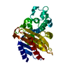
- PDB-1vas: ATOMIC MODEL OF A PYRIMIDINE DIMER SPECIFIC EXCISION REPAIR ENZYM... -
+ Open data

- Basic information
| Entry | Database: PDB / ID: 1vas | ||||||
|---|---|---|---|---|---|---|---|
| Title | ATOMIC MODEL OF A PYRIMIDINE DIMER SPECIFIC EXCISION REPAIR ENZYME COMPLEXED WITH A DNA SUBSTRATE: STRUCTURAL BASIS FOR DAMAGED DNA RECOGNITION | ||||||
 Components Components |
| ||||||
 Keywords Keywords | HYDROLASE/DNA / PROTEIN-DNA COMPLEX / DOUBLE HELIX / HYDROLASE-DNA COMPLEX | ||||||
| Function / homology |  Function and homology information Function and homology informationdeoxyribodipyrimidine endonucleosidase / pyrimidine dimer DNA N-glycosylase activity / deoxyribodipyrimidine endonucleosidase activity / DNA-(apurinic or apyrimidinic site) endonuclease activity / class I DNA-(apurinic or apyrimidinic site) endonuclease activity / DNA-(apurinic or apyrimidinic site) lyase / DNA repair Similarity search - Function | ||||||
| Biological species |  Enterobacteria phage T4 (virus) Enterobacteria phage T4 (virus) | ||||||
| Method |  X-RAY DIFFRACTION / Resolution: 2.75 Å X-RAY DIFFRACTION / Resolution: 2.75 Å | ||||||
 Authors Authors | Vassylyev, D.G. / Kashiwagi, T. / Mikami, Y. / Ariyoshi, M. / Iwai, S. / Ohtsuka, E. / Morikawa, K. | ||||||
 Citation Citation | Journal: Cell(Cambridge,Mass.) / Year: 1995 Title: Atomic model of a pyrimidine dimer excision repair enzyme complexed with a DNA substrate: structural basis for damaged DNA recognition. Authors: Vassylyev, D.G. / Kashiwagi, T. / Mikami, Y. / Ariyoshi, M. / Iwai, S. / Ohtsuka, E. / Morikawa, K. #1: Journal: J.Mol.Biol. / Year: 1995Title: Crystal Structure of a Pyrimidine Dimer-Specific Excision Repair Enzyme from Bacteriophage T4: Refinement at 1.45 Angstroms and X-Ray Analysis of the Three Active Site Mutants Authors: Morikawa, K. / Ariyoshi, M. / Vassylyev, D.G. / Matsumoto, O. / Katayanagi, K. / Ohtsuka, E. #3: Journal: Proc.Natl.Acad.Sci.USA / Year: 1992Title: Role of the Basic Amino Acid Cluster and Glu-23 in Pyrimidine Dimer Glycosylase Activity of T4 Endonuclease V Authors: Doi, T. / Recktenwald, A. / Karaki, Y. / Kikuchi, M. / Morikawa, K. / Ikehara, M. / Inaoka, T. / Hori, N. / Ohtsuka, E. #4: Journal: Science / Year: 1992Title: X-Ray Structure of T4 Endonuclease V: An Excision Repair Enzyme Specific for a Pyrimidine Dimer Authors: Morikawa, K. / Matsumoto, O. / Tsujimoto, M. / Katayanagi, K. / Ariyoshi, M. / Doi, T. / Ikehara, M. / Inaoka, T. / Ohtsuka, E. #5: Journal: J.Mol.Biol. / Year: 1988Title: Preliminary Crystallographic Study of Pyrimidine Dimer-Specific Excision Repair Enzyme from Bacteriophage T4 Authors: Morikawa, K. / Tsujimoto, M. / Ikehara, M. / Inaoka, T. / Ohtsuka, E. | ||||||
| History |
|
- Structure visualization
| Structure viewer | Molecule: MolmilJmol/JSmol |
|---|
- Downloads & links
-Download
| PDBx/mmCIF format | 1vas.cif.gz | 60 KB | Display | PDBx/mmCIF format |
|---|---|---|---|---|
| PDB format | pdb1vas.ent.gz | 40.9 KB | Display | PDB format |
| PDBx/mmJSON format | 1vas.json.gz | Tree view | PDBx/mmJSON format | |
| Others |  Other downloads Other downloads |
-Validation report
| Arichive directory | https://data.pdbj.org/pub/pdb/validation_reports/va/1vasftp://data.pdbj.org/pub/pdb/validation_reports/va/1vas | HTTPS FTP |
|---|
-Related structure data
| Similar structure data |
|---|


-Links
 PDBj
PDBj


- Assembly
| Deposited unit | 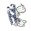
| ||||||||
|---|---|---|---|---|---|---|---|---|---|
| 1 |
| ||||||||
| Unit cell |
|
-Components
| #1: DNA chain | Mass: 3958.571 Da / Num. of mol.: 1 / Source method: obtained synthetically |
|---|---|
| #2: DNA chain | Mass: 3985.613 Da / Num. of mol.: 1 / Source method: obtained synthetically |
| #3: Protein | Mass: 15972.392 Da / Num. of mol.: 1 Source method: isolated from a genetically manipulated source Source: (gene. exp.) Enterobacteria phage T4 (virus) / Genus: T4-like viruses / Species: Enterobacteria phage T4 sensu lato / Gene: DENV / Gene (production host): DENV / Production host:  Keywords: MUTANT E23Q / References: UniProt: P04418 Keywords: MUTANT E23Q / References: UniProt: P04418 |
| #4: Water | ChemComp-HOH /  Mass: 18.015 Da / Num. of mol.: 143 / Source method: isolated from a natural source / Formula: H2O Mass: 18.015 Da / Num. of mol.: 143 / Source method: isolated from a natural source / Formula: H2O |
-Experimental details
-Experiment
| Experiment | Method: X-RAY DIFFRACTION / Number of used crystals: 1 |
|---|
- Sample preparation
| Crystal | Density Matthews: 3.09 Å3/Da / Density % sol: 60.21 % | ||||||||||||||||||||||||||||||||||||
|---|---|---|---|---|---|---|---|---|---|---|---|---|---|---|---|---|---|---|---|---|---|---|---|---|---|---|---|---|---|---|---|---|---|---|---|---|---|
| Crystal grow | Temperature: 277 K / Method: vapor diffusion, hanging drop / pH: 7 Details: pH 7.00, VAPOR DIFFUSION, HANGING DROP, temperature 277.00K | ||||||||||||||||||||||||||||||||||||
| Components of the solutions |
| ||||||||||||||||||||||||||||||||||||
| Crystal | *PLUS | ||||||||||||||||||||||||||||||||||||
| Crystal grow | *PLUS Temperature: 4 ℃ / pH: 7 | ||||||||||||||||||||||||||||||||||||
| Components of the solutions | *PLUS
|
-Data collection
| Diffraction | Mean temperature: 293 K |
|---|---|
| Diffraction source | Source: ROTATING ANODE / Wavelength: 1.5418 Å |
| Detector | Type: MACSCIENCE DIP100 / Detector: IMAGE PLATE / Date: Jun 3, 1994 |
| Radiation | Monochromator: GRAPHITE / Protocol: SINGLE WAVELENGTH / Monochromatic (M) / Laue (L): M / Scattering type: x-ray |
| Radiation wavelength | Wavelength: 1.5418 Å / Relative weight: 1 |
| Reflection | Resolution: 2.75→50 Å / Num. obs: 5662 / % possible obs: 72 % / Observed criterion σ(F): 2 / Redundancy: 6 % / Rmerge(I) obs: 0.062 |
| Reflection | *PLUS Highest resolution: 2.75 Å / Lowest resolution: 50 Å / % possible obs: 72 % / Observed criterion σ(F): 2 / Num. measured all: 24907 |
- Processing
| Software |
| ||||||||||||||||
|---|---|---|---|---|---|---|---|---|---|---|---|---|---|---|---|---|---|
| Refinement | Resolution: 2.75→10 Å / σ(F): 2
| ||||||||||||||||
| Displacement parameters | Biso mean: 7.8 Å2 | ||||||||||||||||
| Refine analyze | Luzzati coordinate error obs: 0.27 Å | ||||||||||||||||
| Refinement step | Cycle: LAST / Resolution: 2.75→10 Å
| ||||||||||||||||
| Software | *PLUS Name: X-PLOR / Version: 3.1 / Classification: refinement | ||||||||||||||||
| Refinement | *PLUS Highest resolution: 2.75 Å / Lowest resolution: 10 Å / Rfactor Rfree: 0.24 | ||||||||||||||||
| Solvent computation | *PLUS | ||||||||||||||||
| Displacement parameters | *PLUS Biso mean: 7.8 Å2 | ||||||||||||||||
| Refine LS restraints | *PLUS
|
