| 登録情報 | データベース: PDB / ID: 1v8g
|
|---|
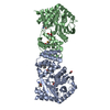
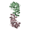
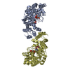
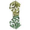
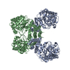
| タイトル | Crystal structure of anthranilate phosphoribosyltransferase (TrpD) from Thermus thermophilus HB8 |
|---|
 要素 要素 | anthranilate phosphoribosyltransferase |
|---|
 キーワード キーワード | TRANSFERASE / Tryptophan biosynthesis / RIKEN Structural Genomics/Proteomics Initiative / RSGI / Structural Genomics |
|---|
| 機能・相同性 |  機能・相同性情報 機能・相同性情報
anthranilate phosphoribosyltransferase / anthranilate phosphoribosyltransferase activity / L-tryptophan biosynthetic process / magnesium ion binding / cytosol類似検索 - 分子機能 Anthranilate phosphoribosyl transferase / Transferase, Pyrimidine Nucleoside Phosphorylase; Chain C / Transferase, Pyrimidine Nucleoside Phosphorylase; Chain A, domain 3 / Pyrimidine Nucleoside Phosphorylase; Chain A, domain 2 / Nucleoside phosphorylase/phosphoribosyltransferase catalytic domain / Glycosyl transferase family 3, N-terminal domain / Glycosyl transferase family 3, N-terminal domain superfamily / Glycosyl transferase family, helical bundle domain / Glycosyl transferase, family 3 / Glycosyl transferase family, a/b domain ...Anthranilate phosphoribosyl transferase / Transferase, Pyrimidine Nucleoside Phosphorylase; Chain C / Transferase, Pyrimidine Nucleoside Phosphorylase; Chain A, domain 3 / Pyrimidine Nucleoside Phosphorylase; Chain A, domain 2 / Nucleoside phosphorylase/phosphoribosyltransferase catalytic domain / Glycosyl transferase family 3, N-terminal domain / Glycosyl transferase family 3, N-terminal domain superfamily / Glycosyl transferase family, helical bundle domain / Glycosyl transferase, family 3 / Glycosyl transferase family, a/b domain / Nucleoside phosphorylase/phosphoribosyltransferase catalytic domain superfamily / Up-down Bundle / 3-Layer(aba) Sandwich / Mainly Alpha / Alpha Beta類似検索 - ドメイン・相同性 |
|---|
| 生物種 |   Thermus thermophilus (バクテリア) Thermus thermophilus (バクテリア) |
|---|
| 手法 |  X線回折 / シンクロトロン / 分子置換 / 解像度: 2.1 Å X線回折 / シンクロトロン / 分子置換 / 解像度: 2.1 Å |
|---|
 データ登録者 データ登録者 | Shimizu, K. / Kunishima, N. / RIKEN Structural Genomics/Proteomics Initiative (RSGI) |
|---|
 引用 引用 | ジャーナル: To be Published
タイトル: Crystal structure of anthranilate phosphoribosyltransferase (TrpD) from Thermus thermophilus HB8
著者: Shimizu, K. / Kunishima, N. |
|---|
| 履歴 | | 登録 | 2004年1月8日 | 登録サイト: PDBJ / 処理サイト: PDBJ |
|---|
| 改定 1.0 | 2004年1月20日 | Provider: repository / タイプ: Initial release |
|---|
| 改定 1.1 | 2008年4月27日 | Group: Version format compliance |
|---|
| 改定 1.2 | 2011年7月13日 | Group: Version format compliance |
|---|
| 改定 1.3 | 2023年10月25日 | Group: Data collection / Database references / Refinement description
カテゴリ: chem_comp_atom / chem_comp_bond ...chem_comp_atom / chem_comp_bond / database_2 / pdbx_initial_refinement_model
Item: _database_2.pdbx_DOI / _database_2.pdbx_database_accession |
|---|
|
|---|
 ムービー
ムービー コントローラー


 データを開く
 基本情報
基本情報 構造の表示
構造の表示 ダウンロードとリンク
 その他のダウンロード


 PDBj
PDBj 集合体
集合体
 分子量: 18.015 Da / 分子数: 543 / 由来タイプ: 天然 / 式: H2O
 試料調製
試料調製 / ビームライン: BL26B2 / 波長: 1 Å
 解析
解析