 Movie
Movie Controller

[English] 日本語
 Yorodumi
- PDB-1v7r: Structure of nucleotide triphosphate pyrophosphatase from pyrococ... -
+ Open data

- Basic information
| Entry | Database: PDB / ID: 1v7r | ||||||
|---|---|---|---|---|---|---|---|
| Title | Structure of nucleotide triphosphate pyrophosphatase from pyrococcus horikoshii OT3 | ||||||
 Components Components | hypothetical protein PH1917 | ||||||
 Keywords Keywords | HYDROLASE / NTPase / STRUCTURAL GENOMICS / RIKEN Structural Genomics/Proteomics Initiative / RSGI | ||||||
| Function / homology |  Function and homology information Function and homology informationpurine nucleoside triphosphate catabolic process / XTP/dITP diphosphatase / ITP diphosphatase activity / XTP diphosphatase activity / dITP diphosphatase activity / nucleotide metabolic process / ribonucleoside triphosphate phosphatase activity / nucleotide binding / metal ion binding / cytoplasm Similarity search - Function | ||||||
| Biological species |   Pyrococcus horikoshii (archaea) Pyrococcus horikoshii (archaea) | ||||||
| Method |  X-RAY DIFFRACTION / SYNCHROTRON / MOLECULAR REPLACEMENT / Resolution: 1.4 Å X-RAY DIFFRACTION / SYNCHROTRON / MOLECULAR REPLACEMENT / Resolution: 1.4 Å | ||||||
 Authors Authors | Lokanath, N.K. / Kunishima, N. / RIKEN Structural Genomics/Proteomics Initiative (RSGI) | ||||||
 Citation Citation | Journal: J.Mol.Biol. / Year: 2008 Title: Structures of dimeric nonstandard nucleotide triphosphate pyrophosphatase from Pyrococcus horikoshii OT3: functional significance of interprotomer conformational changes. Authors: Lokanath, N.K. / Pampa, K.J. / Takio, K. / Kunishima, N. | ||||||
| History |
|
- Structure visualization
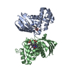
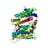
| Structure viewer | Molecule: MolmilJmol/JSmol |
|---|
- Downloads & links
-Download
| PDBx/mmCIF format | 1v7r.cif.gz | 55.2 KB | Display | PDBx/mmCIF format |
|---|---|---|---|---|
| PDB format | pdb1v7r.ent.gz | 39 KB | Display | PDB format |
| PDBx/mmJSON format | 1v7r.json.gz | Tree view | PDBx/mmJSON format | |
| Others |  Other downloads Other downloads |
-Validation report
| Summary document | 1v7r_validation.pdf.gz | 440.9 KB | Display | wwPDB validaton report |
|---|---|---|---|---|
| Full document | 1v7r_full_validation.pdf.gz | 442.1 KB | Display | |
| Data in XML | 1v7r_validation.xml.gz | 11.4 KB | Display | |
| Data in CIF | 1v7r_validation.cif.gz | 16.3 KB | Display | |
| Arichive directory | https://data.pdbj.org/pub/pdb/validation_reports/v7/1v7rftp://data.pdbj.org/pub/pdb/validation_reports/v7/1v7r | HTTPS FTP |
-Related structure data
| Related structure data | 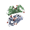 2dvnC  2dvoC  2dvpC 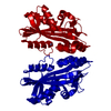 2ztiC 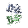 1b78S S: Starting model for refinement C: citing same article ( |
|---|---|
| Similar structure data | |
| Other databases |


-Links
 PDBj
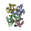
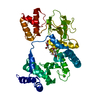
PDBj- Assembly
| Deposited unit | 
| ||||||||
|---|---|---|---|---|---|---|---|---|---|
| 1 | 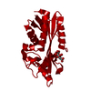
| ||||||||
| Unit cell |
|
-Components
| #1: Protein | Mass: 21233.420 Da / Num. of mol.: 1 Source method: isolated from a genetically manipulated source Source: (gene. exp.) Pyrococcus horikoshii (archaea) / Strain: OT3 / Gene: NTPase / Plasmid: PET-11A / Species (production host): Escherichia coli / Production host:  References: UniProt: O59580, nucleoside-triphosphate diphosphatase |
|---|---|
| #2: Chemical | ChemComp-CIT /   Mass: 192.124 Da / Num. of mol.: 1 / Source method: obtained synthetically / Formula: C6H8O7 Mass: 192.124 Da / Num. of mol.: 1 / Source method: obtained synthetically / Formula: C6H8O7 |
| #3: Water | ChemComp-HOH /  Mass: 18.015 Da / Num. of mol.: 202 / Source method: isolated from a natural source / Formula: H2O Mass: 18.015 Da / Num. of mol.: 202 / Source method: isolated from a natural source / Formula: H2O |
-Experimental details
-Experiment
| Experiment | Method: X-RAY DIFFRACTION / Number of used crystals: 1 |
|---|
- Sample preparation
| Crystal | Density Matthews: 2.41 Å3/Da / Density % sol: 48.61 % | ||||||||||||||||||
|---|---|---|---|---|---|---|---|---|---|---|---|---|---|---|---|---|---|---|---|
| Crystal grow | Temperature: 295 K / Method: microbatch / pH: 5.1 Details: 1.6M Tris sodium citrate, pH 5.1, Microbatch, temperature 295K | ||||||||||||||||||
| Crystal grow | *PLUS Temperature: 291 K | ||||||||||||||||||
| Components of the solutions | *PLUS
|
-Data collection
| Diffraction | Mean temperature: 100 K |
|---|---|
| Diffraction source | Source: SYNCHROTRON / Site: SPring-8  / Beamline: BL26B2 / Wavelength: 1 Å / Beamline: BL26B2 / Wavelength: 1 Å |
| Detector | Type: RIGAKU RAXIS / Detector: IMAGE PLATE / Date: Nov 12, 2003 |
| Radiation | Monochromator: GRAPHITE / Protocol: SINGLE WAVELENGTH / Monochromatic (M) / Laue (L): M / Scattering type: x-ray |
| Radiation wavelength | Wavelength: 1 Å / Relative weight: 1 |
| Reflection | Resolution: 1.4→40 Å / Num. all: 40686 / Num. obs: 40654 / % possible obs: 99.6 % / Observed criterion σ(F): 0 / Observed criterion σ(I): 0 / Redundancy: 7.1 % / Biso Wilson estimate: 17.5 Å2 / Rmerge(I) obs: 0.05 |
| Reflection shell | Resolution: 1.4→1.45 Å / % possible all: 100 |
| Reflection | *PLUS Num. obs: 40686 / % possible obs: 99.8 % / Num. measured all: 286969 / Rmerge(I) obs: 0.05 |
| Reflection shell | *PLUS % possible obs: 100 % / Rmerge(I) obs: 0.272 / Mean I/σ(I) obs: 5.2 |
- Processing
| Software |
| ||||||||||||||||||||||||||||||||||||
|---|---|---|---|---|---|---|---|---|---|---|---|---|---|---|---|---|---|---|---|---|---|---|---|---|---|---|---|---|---|---|---|---|---|---|---|---|---|
| Refinement | Method to determine structure: MOLECULAR REPLACEMENT Starting model: 1B78 Resolution: 1.4→26.5 Å / Rfactor Rfree error: 0.005 / Data cutoff high absF: 1374304.66 / Data cutoff low absF: 0 / Cross valid method: THROUGHOUT / σ(F): 0 / Stereochemistry target values: Engh & Huber
| ||||||||||||||||||||||||||||||||||||
| Solvent computation | Solvent model: FLAT MODEL / Bsol: 42.463 Å2 / ksol: 0.38057 e/Å3 | ||||||||||||||||||||||||||||||||||||
| Displacement parameters | Biso mean: 18.5 Å2
| ||||||||||||||||||||||||||||||||||||
| Refine analyze |
| ||||||||||||||||||||||||||||||||||||
| Refinement step | Cycle: LAST / Resolution: 1.4→26.5 Å
| ||||||||||||||||||||||||||||||||||||
| Refine LS restraints |
| ||||||||||||||||||||||||||||||||||||
| LS refinement shell | Highest resolution: 1.4 Å / Total num. of bins used: 6 /
| ||||||||||||||||||||||||||||||||||||
| Xplor file |
| ||||||||||||||||||||||||||||||||||||
| Refinement | *PLUS Lowest resolution: 40 Å / Rfactor Rfree: 0.222 | ||||||||||||||||||||||||||||||||||||
| Solvent computation | *PLUS | ||||||||||||||||||||||||||||||||||||
| Displacement parameters | *PLUS | ||||||||||||||||||||||||||||||||||||
| Refine LS restraints | *PLUS
|
