Entry Database : PDB / ID : 1uyoTitle Translocator domain of autotransporter NalP from Neisseria meningitidis NALP Keywords / / / / / Function / homology Function Domain/homology Component
/ / / / / / / / / / / / / / / / / / / / / / / / / / / / / / / / / / Biological species NEISSERIA MENINGITIDIS (bacteria)Method / / / Resolution : 3.2 Å Authors Oomen, C.J. / Van Ulsen, P. / Van Gelder, P. / Feijen, M. / Tommassen, J. / Gros, P. Journal : Embo J. / Year : 2004Title : Structure of the Translocator Domain of a Bacterial AutotransporterAuthors : Oomen, C.J. / Van Ulsen, P. / Van Gelder, P. / Feijen, M. / Tommassen, J. / Gros, P. History Deposition Mar 2, 2004 Deposition site / Processing site Revision 1.0 Mar 19, 2004 Provider / Type Revision 1.1 May 7, 2011 Group Revision 1.2 Jul 13, 2011 Group Revision 1.3 Jul 24, 2019 Group / Derived calculations / Category / struct_connItem / _struct_conn.pdbx_leaving_atom_flagRevision 1.4 Oct 16, 2024 Group Data collection / Database references ... Data collection / Database references / Other / Structure summary Category chem_comp_atom / chem_comp_bond ... chem_comp_atom / chem_comp_bond / database_2 / pdbx_database_status / pdbx_entry_details / pdbx_modification_feature Item / _database_2.pdbx_database_accession / _pdbx_database_status.status_code_sf
Show all Show less Remark 700 SHEET DETERMINATION METHOD: AUTHOR PROVIDED.
 Movie
Movie Controller

 Yorodumi
 Open data
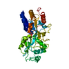
 Basic information
 Components
 Keywords
 Function and homology information
 NEISSERIA MENINGITIDIS (bacteria)
 X-RAY DIFFRACTION /
  Authors
 Citation
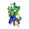
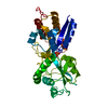
 Structure visualization
 Downloads & links
 Other downloads
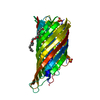


 PDBj
PDBj


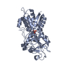
 Assembly
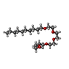
 Mass: 378.544 Da / Num. of mol.: 1 / Source method: obtained synthetically / Formula: C20H42O6
 Sample preparation
 / Beamline: X11 / Wavelength: 0.811
  Processing